Showing posts with label Top Medical Conferences. Show all posts

Thursday, 10 January 2019

Genetics may influence the effects of vitamin E on cancer risk


Almost half of all Americans take a vitamin supplement, and yet many large-scale, placebo-controlled clinical trials of various supplements have found little or no benefit. A new study led by investigators from Brigham and Women's Hospital suggests an intriguing reason for this: genetic variation may be influencing these effects, increasing risk in some individuals while decreasing risk for others.

Investigators conducted a retrospective analysis of the Women's Health Study (WHS) and its genetic component, the Women's Genome Health Study (WGHS), as well as validated their results in the Alpha-Tocopherol Beta-Carotene Cancer Prevention Study (ATBC). Both of these trials investigated whether taking vitamin E supplements could affect risk of cancer. They found that genetic variations in the gene COMT influenced whether vitamin E decreased or increased risk of developing cancer during and after the study periods. Their results are published online in The Journal of the National Cancer Institute.
"Observational studies of people taking vitamin E have reported benefits, and studies in animal models have suggested a protective effect, but when vitamin E supplements were brought into placebo-controlled clinical trials, the results were null," said Kathryn Hall, PhD, MPH, from the Division of Preventive Medicine at the Brigham. "This made it easy to assume that vitamin E just doesn't work. But what we've found is that it may have been protective in some and not in others, and that genetic variation is linked to these outcomes."
Multiple pathways may link the enzyme catechol-O-methyltransferase (COMT) to how vitamin E is processed by the body. Hall and colleagues previously found that genetic differences in COMT modified the effects of vitamin E on cardiovascular disease risk.

The most extensively studied variant in COMT comes in three genetic "flavors": met/met, val/met and val/val. People with the val/val variant have a version of COMT that is 3-4 times more enzymatically active than people with met/met.

Hall and her co-authors, including colleagues from the Division of Preventive Medicine -- Nancy Cook, ScD, Julie Buring, ScD, Howard Sesso, ScD, and Daniel Chasman, PhD -- as well as Paul Ridker, MD, director of the Center for Cardiovascular Disease Prevention, looked at cancer rates among women during the WHS/WGHS trial, which lasted 10 years, plus the 10 years immediately following the trial's conclusion. They found that in the overall period, for women with the met-met variant who took vitamin E compared to placebo, rates of total cancer were 14 percent significantly lower, while they were 15 percent higher for women with the val-val variant who took vitamin E compared to placebo.

Participants in the WGHS were all female health care professionals in good health at the beginning of the trial. The researchers validated these results by looking at a second cohort. Similar results were seen in the ATBC trial, led by Demetrius Albanes, MD, MPH, of the National Institutes of Health, which included only male smokers from Finland.

The team also analyzed rates of cancer sub-types such as breast, lung, uterine and colorectal cancer, finding similar trends to the overall cancer rates.
"Significant gene-drug interactions are hard to find, and this one is particularly striking. Now we need to understand which cancers are affected, why and how, and these results encourage us to pursue this with robust and rigorous curiosity," said Hall.
Researchers from different part of the world are invited to submit abstract on their unpublished latest research at our upcoming conference Cell Tissue Science 2019 which is focused on the complications and consequences of Stem CellRegenerative MedicineStem Cell TherapyCancer Cell Biology,Technical Advancements in cancer treatmentand many more. We as committee members of the conference welcome you to be a part of the conference “ 12th World Congress on Cell & Tissue Science” in Singapore on March 11-12, 2019
You can submit your abstract on Session or Track : 08. Advancement in Cancer Treatments
With Regards to Christmas and New Year Celebration we are providing a special discount of 30% on all Registration Categories for more information please  visit by Click Here

Wednesday, 9 January 2019

New mechanism to 'activate' the immune system against cancer


According to a study published this week in the journal Nature, A new mechanism for activating the immune system against cancer cells allows immune cells to detect and destroy cancer cells better than before,

The focus of the study is a mechanism that routinely serves the cell by marking human virus-like genes in order to avoid identifying them as viruses. Now, Prof. Levanon, together with the Harvard team, has discovered that when inhibiting this mechanism, the immune system can be harnessed to fight cancer cells in a particularly efficient manner, and most effectively in lung cancer and melanoma.
"We found that if the mechanism is blocked, the immune system is much more sensitive. When the mechanism is deactivated, the immune system becomes much more aggressive against the tumor cells," said Prof. Erez Levanon, doctoral student Ilana Buchumansky of the Mina and Everard Goodman Faculty of Life Sciences, Bar-Ilan University
In recent years, a new generation of cancer drugs has been developed which blocks proteins that inhibit immune activity against malignant tumors. These drugs have shown remarkable success in several tumor types. This year's Nobel Prize in Medicine was awarded to James Allison and Tasuku Honjo, who discovered the key genes of this mechanism. Despite this achievement, the current generation of drugs helps only a small number of patients, while most of the drugs fail to cause the immune system to attack the tumor. It is hoped that the new discovery will allow enhanced activity of the immune system to attack cancer cells. A number of companies have already begun research to screen for drugs that will operate on the basis of this discovery.
Researchers from different part of the world are invited to submit abstract on their unpublished latest research at our upcoming conference Cell Tissue Science 2019 which is focused on the complications and consequences of Stem CellRegenerative MedicineStem Cell TherapyCancer Cell Biology,Technical Advancements in cancer treatment and many more. We as committee members of the conference welcome you to be a part of the conference “ 12th World Congress on Cell & Tissue Science” in Singapore on March 11-12, 2019
You can submit your abstract on Session or Track : 08. Advancement in Cancer Treatments
With Regards to Christmas and New Year Celebration we are providing a special discount of 30% on all Registration Categories for more information please  visit by Click Here
Saturday, 13 October 2018

Bug that causes stomach cancer could play a role in colorectal cancer

A bacterium known for causing stomach cancer might also increase the risk of certain colorectal cancers, particularly among African Americans, according to a study led by Duke Cancer Institute researchers.

The finding describes an association between antibodies to H. pylori bacteria and an increased risk of colorectal cancers, although it does not establish the bacteria as a definitive cause; those studies are ongoing.

But in an analysis of more than 4,000 colorectal cancer cases culled from large, diverse cohort studies, the researchers found a significant correlation between colorectal cancer incidence and those who had been infected with a virulent strain of H. pylori that is especially common among African Americans.
"The link between infection and cancer is intriguing, particularly if we can eradicate it with a simple round of antibiotics," said Meira Epplein, Ph.D., Co-leader of Cancer Control and Population Sciences at Duke Cancer Institute. "Our study provides strong evidence that we need to pursue this research to establish a definitive cause-and-effect."
They analyzed blood samples from more than 8,400 ethnically and regionally diverse study participants -- half who went on to develop colorectal cancer and the other half with no such diagnosis.

The researchers found that H. pylori infections were equally common in both the cancer and non-cancer group, with 4 in 10 patients in both groups testing positive for exposure to the bacterium.

But stark racial differences also appeared. White patients had below average H. pylori infection rates, and Asian Americans had average rates. For black and Latino patients, however, the rates were much higher. Among African Americans, 65 percent of the non-cancer patients and 71 percent of the colorectal cancer patients had H. pylori antibodies; among Latinos, 77 percent of the non-cancer group and 74 percent of the cancer group had antibodies.

Further analysis showed that antibodies to four H. pylori proteins were most often present among the different ethnic groups with colorectal cancer. One H. pylori protein in particular, VacA, had the strongest association with increased odds of colorectal cancer among the African American patients in the study, and, specifically, high levels of antibodies to this protein were associated with colorectal cancer incidence in both African Americans and Asian Americans.
"It was surprising to find VacA antibodies increased the odds of colorectal cancer in African Americans and Asian Americans, and not in whites and Latinos," Epplein said. "This is a big question -- are people harboring different bacteria based on genetic origin or heritage? This is part of what we need to figure out."
Epplein said additional studies might also determine whether antibodies to the H. pylori VacA protein could serve as a marker of colorectal cancer risk if it isn't causing the cancer directly.
We welcome researchers from different part of the to submit abstract on their latest research at our upcoming conference Cell Tissue Science 2019 which is mainly focuses on the complications the consequences of Stem CellRegenerative MedicineStem Cell TherapyCancer Cell Biology , Technical Advancements in cancer treatment and many more.We welcome you to the our upcoming conference “ 12th World Congress on Cell & Tissue Science” . For more info visit :Cell Tissue Science 2019

Friday, 12 October 2018

Redrawing the structure of an immune system protein

Researchers have revealed the structure of an essential immune protein, creating future possibilities to develop more effective medicines for a range of illnesses from cancer to neurological diseases. University of Tokyo researchers made this discovery with computerized image analysis and modern electron microscope imaging.
Cell Tissue Science2019
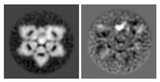
Electron microscopes and modern computer image processing allowed University of Tokyo researchers to uncover the correct structure of a protein essential in the immune system, IgM, and a much smaller protein that is bound inside, AIM. IgM is the larger, incomplete hexagon shape and AIM is the smaller broad bean shape inside the wedge-shape gap. In the right-side image, AIM is more easily recognized as the bright white, broad-bean-shaped spot.
Credit: Image by Hiramoto et al., originally published in Science Advances, CC-BY
Researchers verified the structure of natural immunoglobulin M (IgM) protein, an important part of the immune system, using mouse and human versions of the protein. IgM is now understood to be shaped like an incomplete hexagon, or a pentagon with a wedge-shaped gap.

IgM is the first immune system protein that develops in the human fetus and remains the first responder to pathogens throughout life. The structure of IgM was first identified in 1969 as a "five-pointed, star-shaped table" and updated in 2009 to be a five-sided dome or "mushroom cap."
"The original IgM model was made by looking at a few individual molecules by hand with what today we think is a low-resolution microscope. Now we have clearer pictures and the computer can examine thousands of individual IgM molecules," said Miyazaki.
The 2018 discovery of the incomplete hexagon was actually a secondary interest for Miyazaki, originally a medical doctor who has built his research career studying a different protein called apoptosis inhibitor of macrophage (AIM).

Since identifying the correct shape of IgM, researchers now understand that inactive AIM is nestled inside the gap of the IgM incomplete hexagon. The structural connection between IgM and AIM means that drugs with the ability to regulate the release of AIM could be used to create AIM-based disease therapies.
"We can think of AIM as a fighter jet and IgM as the aircraft carrier ship. When other molecules activate the immune system, IgM releases AIM. The much smaller AIM protein goes around the body to clear away damaged cells and prevent disease," said Miyazaki.
Miyazaki identified AIM in 1999 while working at the Basel Institute for Immunology Institute in Switzerland. Its small size means AIM is easily eliminated from the body by the kidneys and excreted in urine, so staying bound within the larger IgM protects AIM from being removed before it is needed.

AIM is a common molecule in the bloodstream, but it is only active when the body develops a disease. AIM is known to be important for preventing obesity, fatty liver disease, hepatocellular carcinoma (liver cancer), multiple sclerosis (MS), fungus-induced peritonitis (inflammation of the abdominal wall membrane), and acute kidney injury.

The incomplete hexagon structure is still only a 2D understanding of IgM structure. Miyazaki and his team continue to do additional analysis and hope to report the 3D structure of IgM soon.
We welcome researchers from different part of the to submit abstract on their latest research at our upcoming conference Cell Tissue Science 2019 which is mainly focuses on the complications the consequences of Stem CellRegenerative MedicineStem Cell TherapyCancer Cell Biology , Technical Advancements in cancer treatment and many more.We welcome you to the our upcoming conference “ 12th World Congress on Cell & Tissue Science” . For more info visit :Cell Tissue Science 2019

Wednesday, 10 October 2018

There's a better way to decipher DNA's epigenetic code to identify disease

A new method for sequencing the chemical groups attached to the surface of DNA is paving the way for better detection of cancer and other diseases in the blood, according to research from the Perelman School of Medicine at the University of Pennsylvania
These chemical groups mark one of the four DNA "letters" in the genome, and it is differences in these marks along DNA that control which genes are expressed or silenced.

Cell Tissue Science 2019
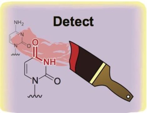
Enzymes, rather than harsh chemical reactions, can be used to reveal the epigenetic code in DNA.
Credit: Rahul Kohli, Univeristy of Pennsylvania

To detect disease earlier and with increased precision, researchers have a growing interest in analyzing free-floating DNA in settings in which there is a limited amount, such as that extruded from tumors into the bloodstream.
"We're hopeful that this method offers the ability to decode epigenetic marks on DNA from small and transient populations of cells that have previously been difficult to study, in order to determine whether the DNA is coming from a specific tissue or even a tumor." said Rahul Kohli, MD, PhD, Assistant Professor of Biochemistry and Biophysics, and Medicine.
For the last several decades, the major methods used to decipher the epigenetic code have relied on a chemical called bisulfite. While bisulfite has proven useful, it also presents major limitations: it is unable to differentiate the most common modifications on the DNA building block cytosine, and more significantly, it destroys much of the DNA it touches, leaving little material to sequence in the lab.

The new method described in this paper builds on the fact that a class of immune-defense enzymes, called APOBEC DNA deaminases, can be repurposed for biotech applications. Specifically, the deaminase-guided chemical reaction is able to achieve what bisulfite could do, but without harming DNA.
"This technological advance paves the way to better understand complex biological processes such as how the nervous system develops or how a tumor progresses," said Hao Wu, PhD, Assistant Professor of Genetics. 
Using this method, the team showed that determining the epigenetic code of one type of neuron used 1,000-times less DNA than required by the bisulfite-dependent methods. From this, the new method could also differentiate between the two most common epigenetic marks, methylation and hydroxmethylation.
"We were able to show that sites along the genome that appear to be modified are in fact very different in terms of the distribution of these two marks," Kohli said. "This finding suggests important and distinctive biological roles for the two marks on the genome."
We encourage researchers all around the globe to submit abstract on their latest research at our upcoming conference Cell Tissue Science 2019 which is mainly focuses on the complications the consequences of Stem CellRegenerative MedicineStem Cell TherapyCancer Cell Biology , Technical Advancements in cancer treatment  ,Epigenetics and many more.We welcome you to the our upcoming conference “ 12th World Congress on Cell & Tissue Science” . For more info visit :Cell Tissue Science 2019

Monday, 8 October 2018

Novel mechanism for generating our skeleton

There are more than 200 bones in the human body. Bone is formed during embryonic and postnatal skeletogenesis by two distinct, well-organized processes, intramembranous and endochondral ossification.

Mesenchymal stem cells differentiate into chondrocytes to form a cartilaginous template, which, for long bones, induces bone formation through endochondral ossification. Extracellular signal-regulated kinase 5 (Erk5), which is a member of the mitogen-activated protein kinase (MAPK) family, is phosphorylated by MAPK/Erk kinase-5 (Mek5) to regulate the function of various cell types.

Although the MAPK Erk1/2 pathway is well known for regulating skeletogenesis, the in vivo physiological role of the Mek5/Erk5 pathway in skeletal development has been largely unclear to date, because of the early embryonic lethality of global Erk5 knockout mice.

181002103035_1_900x600
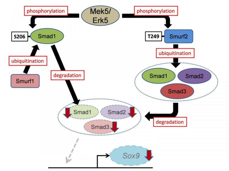
Schematic model of this study.


By using cell-specific mouse genetics approaches revealed that Erk5 plays a crucial role in skeletogenesis in vivo. Paired-related homeobox 1 (Prx1) is expressed in mesenchymal stem cells in the limb buds. Mesenchymal stem cell-specific Erk5 knockout embryos (Prx1-Cre;Erk5fl/fl embryos) displayed wider long bones and impaired mineralization of the metatarsal. In contrast, chondrocyte-specific Erk5 knockout embryos (Col2a1-Cre;Erk5fl/fl embryos) recapitulated only the wider long bone phenotype of Prx1-Cre;Erk5fl/fl embryos. Accordingly, the investigators revealed that Erk5 controls.
  1. Early chondrogenic differentiation of mesenchymal stem cells, including mesenchymal condensation, through its expression in skeletogenic progenitors
  2. Chondrogenic differentiation after the formation of mesenchymal condensations through its expression in chondrocytes.
Subsequent biochemical analyses found that Erk5 directly phosphorylates Smad1 at Ser206 in the linker region, which is known to trigger its proteasomal degradation in a Smad-specific E3 ubiquitin ligase 1 (Smurf1)-dependent manner. In addition, Erk5 directly phosphorylates Smurf2 at Thr249, leading to accelerated proteasomal degradation of Smad proteins (Smad1, 2 and 3).

The study demonstrated that Smads transcriptionally activate the expression in mesenchymal stem cells of sex-determining region within the Y-type high-mobility group box protein 9 (Sox9), which is the principal transcription factor involved in skeletogenesis. Moreover, using mouse genetic rescue experiments, the investigators revealed that Sox9 is a critical mediator of Erk5-dependent skeletogenesis.

 In conclusion, the Mek5/Erk5 pathway is critical for skeletogenesis in vivo through its expression in mesenchymal stem cells and modulation of Smad protein stability (Smad1, 2, and 3) via Smurf activity (Smurf1 and 2). These findings improve our understanding of the molecular mechanisms underlying skeletal development and spur the development of drugs targeting human cartilage diseases associated with abnormal chondrocyte differentiation and maturation. Moreover, since the Smurf2/Smads cascade is associated with other diseases, including cancer and ageing, the newly identified Mek5/Erk5/Smurf2/Smads/Sox9 cascade is a candidate target for developing drugs to treat a variety of human diseases.
 
We welcome researchers from different part of the to submit abstract on their latest research at our upcoming conference Cell Tissue Science 2019 which is mainly focuses on the complications the consequences of Stem CellRegenerative MedicineStem Cell TherapyCancer Cell Biology , Technical Advancements in cancer treatment and many more.We welcome you to the our upcoming conference “ 12th World Congress on Cell & Tissue Science” . For more info visit :Cell Tissue Science 2019

Thursday, 4 October 2018

Diet affects the breast microbiome in mammals

Diet influences the composition of microbial populations in the mammary glands of nonhuman primates. Specifically, a Mediterranean diet increased the abundance of probiotic bacteria previously shown to inhibit tumor growth in animals.
Cell Tissue Science 2019
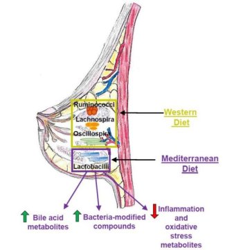
This image shows how diet plays a critical role in determining microbiota populations in tissues outside the gut, such as the mammary gland. Credit: Katherine Cook

"We showed for the first time that breast-specific microbiome populations are significantly affected by diet, and this was in a well-established nonhuman primate model of women's health, increasing the likelihood that these findings will be important for human health," says Carol Shively, Wake Forest School of Medicine. "The breast microbiome is now a target for intervention to protect women from breast cancer."
Diet has been extensively studied as a lifestyle factor that could influence breast cancer development. Breast cancer risk in women is increased by consumption of a high-fat Western diet full of sweets and processed foods but reduced by a healthy Mediterranean diet consisting of vegetables, fish, and olive oil. Intriguingly, a recent study in humans revealed that malignant breast tumors have a lower abundance of Lactobacillus bacteria compared to benign lesions, suggesting that microbial imbalances could contribute to breast cancer.
"However, it was unknown what possible factors could modulate the breast tissue microbiome," says Katherine Cook, Wake Forest School of Medicine. "Diet is a strong influencer on the gut microbiome, so we decided to test the hypothesis that diet can impact mammary gland microbiota populations."
To address this question, Shively and Cook used macaque monkeys because the animals mimic human breast biology and have been used to study breast cancer risk. One advantage over human studies is that the food intake of the monkeys can be carefully controlled for a prolonged period of time, increasing the chance of observing profound effects of diet.
The researchers assigned 40 adult female monkeys to receive either a Western or a Mediterranean diet for 31 months. The breast tissue of monkeys that consumed a Mediterranean diet had a 10-fold higher abundance of Lactobacillus, which is commonly used in probiotics and has been shown to decrease tumor growth in animals with breast cancer. The Mediterranean diet also increased levels of bile acid metabolites and bacterial-processed bioactive compounds that may decrease breast cancer risk.
Taken together, these results suggest that diet directly influences microbiome populations outside of the intestinal tract and could impact mammary gland health. But for now, it is not clear what impact these microbes or microbial-modified metabolites have on breast cancer risk.
Moving forward, the Cook lab plans to investigate the physiological impact of increased Lactobacillus in mammary gland tissue. They will also assess whether oral interventions such as fish oil or probiotic supplements can impact microbial populations in mammary glands and breast tumors. In addition, they are exploring the role of bacterial-modified bioactive compounds and bile acids on inflammation, breast cancer tumor growth, and therapeutic responsiveness.
"Our future studies are designed to validate the use of probiotics, fish oil, or antibiotics during neoadjuvant therapy to improve therapeutic outcomes," Cook says. "We hope to translate our studies into the clinic in the near future."
We encourage researchers all around the globe to submit abstract on their latest research at our upcoming conference Cell Tissue Science 2019 which is mainly focuses on the complications the consequences of Stem CellRegenerative MedicineStem Cell TherapyCancer Cell Biology , Technical Advancements in cancer treatment   and many more.We welcome you to the our upcoming conference “ 12th World Congress on Cell & Tissue Science” . For more info visit :Cell Tissue Science 2019