Showing posts with label Stem Cell Conference. Show all posts

Friday, 7 June 2019

New genes out of nothing


One key question in evolutionary biology is how novel genes arise and develop. Swedish researchers now show how new genes and functions that are advantageous to bacteria can be selected from random DNA sequences. The results are presented in the scientific journal mBio.

How do new genes and functional proteins arise and develop? This is one of the most fundamental issues in evolutionary biology. Two different types of mechanism have been proposed: (1) new genes with novel functions arise from existing genes, and (2) new genes and proteins evolve from random DNA sequences with no similarity to existing genes and proteins.

In the present study, the researchers explored the latter type of mechanism: evolution of new genes and proteins from randomised DNA sequences as its called as de novo evolution. It is fairly easy to understand that when a gene already exists, it can be modified and acquire a new function. But how does "nothing" turn into a function affording a small advantage that is favoured by natural selection?

The raw material for the experiment was an big library of some 500 million randomised gene sequences, from which peptide sequences with a biological function were identified. In the experiment, random gene sequences were placed on a plasmid and overexpressed. The scientists then investigated whether they could give bacteria a specific, defined property. Were they, for example, able to give the bacteria antibiotic resistance? They identified several short peptides (22-25 amino acids long) that could give the bacteria a high degree of resistance to aminoglycosides, an important class of antibiotics used for severe infections.
"When the project started, we had low expectations. We were amazed when we found peptides able to confer a resistance level 48 times higher," says Dr Michael Knopp, the study's lead author.
Through a combination of genetic and functional experiments, the scientists were able to demonstrate that the peptides cause resistance by attaching themselves to bacterial cell membranes and affecting the proton potential across the membrane. The disruption of the proton potential causes a decrease in antibiotic uptake, rendering the bacteria resistant.
"This study is important because it shows that completely random sequences of amino acids can give rise to new, advantageous functions, and that this process of de novo evolution can be studied experimentally in the laboratory," says Dan I. Andersson, Professor of Medical Bacteriology, who is chiefly responsible for the study.
An important announcement regarding our upcoming conference 12th World Congress on Cell & Tissue Science (Cell Tissue Science 2019) scheduled on September 13-14,2019 in Singapore. You can also present your latest research at the different topics such as Cancer Cell Biology, Stem Cell & its applications and many more along with other distinguished professors, doctors and researchers from all over the world.
If interested kindly proceed with submitting your abstract and latest biography along with a photography to our online abstract submission page given below: Link for submission: Click Here
Source of the article: https://www.sciencedaily.com/releases/2019/06/190604131214.htm

Saturday, 1 June 2019

How prostate cancer cells mimic bone when they metastasize


Prostate cancer often becomes lethal as it spreads to the bones, and the process behind this deadly feature could potentially be turned against it as a target for bone-targeting radiation and potential new therapies.

Study published online in the journal PLOS ONE, Duke Cancer Institute researchers describe how prostate cancer cells develop the ability to mimic bone-forming cells called osteoblasts, enabling them to proliferate in the bone microenvironment.

Attacking these cells with radium-233, a radioactive isotope that selectively targets cells in these bone metastases, has been shown to prolong patients' lives. But a better understanding of how radium works in the bone was needed.

The mapping of this mimicking process could lead to a more effective use of radium-233 and to the development of new therapies to treat or prevent the spread of prostate cancer to bone.
"Given that most men who die of prostate cancer have bone metastases, this work is critical to helping understand this process," said lead author Andrew Armstrong, Director of Research at the Duke Cancer Institute Center for Prostate and Urologic Cancers.
The research team enrolled a small study group of 20 men with symptomatic bone-metastatic prostate cancer. When analyzing the circulating tumor cells from study participants, they found that bone-forming enzymes appeared to be expressed commonly, and that genetic alterations in bone forming pathways were also common in these prostate cancer cells.

They validated these new genetic findings in a separate multicenter trial involving a larger group of more than 40 men with prostate cancer and bone metastases.

Following treatment with radium-223, the researchers found that the radioactive isotope was concentrated in bone metastases, but tumor cells still circulated and cancer progressed within six months of therapy. The researchers found a range of complex genetic alterations in these tumor cells that likely enabled them to persist and develop resistance to the radiation over time.
"Osteomimicry may contribute in part to how prostate cancer spreads to bone, but also to the uptake of radium-223 within bone metastases and may thereby enhance the therapeutic benefit of this bone targeting radiotherapy," Armstrong said.
He said by mapping this lethal pathway of prostate cancer bone metastasis, the study points to new targets and thus critical areas of research into designing better tumor-targeting therapies.
An important announcement regarding our upcoming conference 12th World Congress on Cell & Tissue Science (Cell Tissue Science 2019) scheduled on September 13-14,2019 in Singapore. You can also present your latest research at the different topics such as Cancer Cell Biology, Stem Cell & its applications and many more along with other distinguished professors, doctors and researchers from all over the world.
If interested kindly proceed with submitting your abstract and latest biography along with a photography to our online abstract submission page given below: Link for submission: Click Here

Saturday, 19 January 2019

How stem cells self-organize in the developing embryo


Embryonic development is a process of profound physical transformation, one that has challenged researchers for centuries. How do genes and molecules control forces and tissue stiffness to orchestrate the emergence of form in the developing embryo? How are the precise mechanics underlying emergence of the complexity of our organs and tissues encoded in our DNA?

One particular aspect of embryonic development: how a group of stem cells -- the endoderm -- moves from the surface of the developing embryo to the center, and in doing so transforms from a flat sheet to a hollow tube. This structure, known as the gut tube, then forms the lining of the entire respiratory and gastrointestinal tracts.

In a study published today in Nature, Nerurkar worked with colleagues at Harvard to shed new light on this critical step in early embryonic development. The team discovered gut tube formation is driven by collective cell movements of the endoderm, a process by which cells travel large distances en masse, without rearranging relative to one another. They also found that this collective movement is triggered by cells that are converting a molecular gradient to a force gradient that drives cells from the surface into the embryo. This discovery is one of the few examples, especially among vertebrates, of how molecular cues are converted into the physical forces that shape our organs.
The study findings could have important implications for how stem cells are used to create functional organs in the lab, and lead to a better understanding of the underlying causes of gastrointestinal birth defects. "Our major goal is to understand how we, as complex organisms, are formed with such precision from a seemingly disorganized ball of cells -- the early embryo," says Nandan Nerurkar, assistant professor of biomedical engineering at Columbia Engineering.
Identifying genes that drive differentiation of stem cells into mature cell types -- the primary focus in Nerurkar's field -- is an important step toward growing replacement organs in the lab. However, Nerurkar suggests this is only part of the picture: "It is equally important to understand how to instruct those cells to organize into functional three-dimensional organs. The developing embryo holds the recipe for this, and many research groups, including ours, are now leveraging the language of physics and mechanics to dissect it."

The team used an innovative approach at the leading edge of the developmental biology field. They combined conventional approaches of developmental biology, including analysis and manipulation of gene expression and live time lapse microscopy of cell movements in the developing chick embryo, with engineering methods, such as mathematical modeling and force and strain measurements.

They focused on one part of endoderm internalization: the hindgut, which gives rise to half of the small intestine, the large intestine, and colon. What was previously known of gut tube formation came from fate-mapping experiments, wherein cells are labeled early in development and then mapped to where the labeled cells end up later in development. This static analysis, which uses static images of the beginning and the end of the process to make an educated guess of what happens in the middle, has led to a view of gut tube formation that is present in most embryology textbooks. "Based on our recent findings, this view is at best incomplete, and at worst completely wrong," says Nerurkar.

Unlike earlier fate-mapping studies, Nerurkar and his colleagues used live imaging in the embryo to directly observe cell movements as the endoderm is internalized to form a tube. They next applied a combination of mechanical engineering and developmental biology approaches to understand just how those cell movements occur, and how the movements are coordinated to form this critical structure in the early embryo.

The team found that the movements are coordinated by the conversion of a molecular gradient into a force gradient from cells that are contracting in proportion to the amount of a molecular cue -- fibroblast growth factor (FGF) -- that they sense. This results in a tug of war among endoderm cells: as one "team" begins to win, the cells actually recruit players from the opposing team by pulling them from low to higher concentrations of FGF.
Irregularities in FGF function can lead to a number of developmental defects. "During human development, errors in gut tube formation would likely lead to miscarriage, something that is a relatively high risk during the first trimester, when this process is occurring," says Nerurkar.
While this study focused on just one part of endoderm internalization, the hindgut, it is still unknown how the foregut, which forms the trachea, lungs, esophagus, stomach, and liver, and the midgut, which forms the pancreas and small intestine, are formed. Nerurkar plans to use his new approach to study these other areas of embryonic development and investigate if and how FGF signaling acts more broadly to control mechanics in the development of other tissues and organs.
"I want to learn more about how mechanics and molecules are integrated to coordinate the formation of these very distinct tissues by disparate mechanisms, yet from the same initial pool of stem cells," he says. "By focusing on the tissue-level mechanics downstream of FGF signaling, we may now be able to understand what this important pathway does to shape other organs and tissues during development, including the heart, brain, and spinal column."
Nerurkar is continuing this research at Columbia Engineering, developing quantitative molecular-mechanical relationships that could be used to design and construct replacement tissues in the lab, using controlled delivery of these diffusible cues -- the instructional signals that are secreted by cells and then float away to neighboring cells -- to instruct the self organization of cells into functional tissues and organs. If he and others in this field can establish the design principles of embryonic tissue formation, it will be possible to repurpose those same principles for regenerative medicine and tissue engineering applications.
Researchers from different part of the world are invited to submit abstract on their unpublished latest research at our upcoming conference Cell Tissue Science 2019 which is focused on the complications and consequences of Stem Cell, Regenerative Medicine, Stem Cell Therapy, Cancer Cell Biology,Technical Advancements in cancer treatment and many more. We as committee members of the conference welcome you to be a part of the conference “ 12th World Congress on Cell & Tissue Science” in Singapore on March 11-12, 2019. 
You can submit your abstract on Session or Track : 09. Stem Cells and its Applications

Wednesday, 9 January 2019

New mechanism to 'activate' the immune system against cancer


According to a study published this week in the journal Nature, A new mechanism for activating the immune system against cancer cells allows immune cells to detect and destroy cancer cells better than before,

The focus of the study is a mechanism that routinely serves the cell by marking human virus-like genes in order to avoid identifying them as viruses. Now, Prof. Levanon, together with the Harvard team, has discovered that when inhibiting this mechanism, the immune system can be harnessed to fight cancer cells in a particularly efficient manner, and most effectively in lung cancer and melanoma.
"We found that if the mechanism is blocked, the immune system is much more sensitive. When the mechanism is deactivated, the immune system becomes much more aggressive against the tumor cells," said Prof. Erez Levanon, doctoral student Ilana Buchumansky of the Mina and Everard Goodman Faculty of Life Sciences, Bar-Ilan University
In recent years, a new generation of cancer drugs has been developed which blocks proteins that inhibit immune activity against malignant tumors. These drugs have shown remarkable success in several tumor types. This year's Nobel Prize in Medicine was awarded to James Allison and Tasuku Honjo, who discovered the key genes of this mechanism. Despite this achievement, the current generation of drugs helps only a small number of patients, while most of the drugs fail to cause the immune system to attack the tumor. It is hoped that the new discovery will allow enhanced activity of the immune system to attack cancer cells. A number of companies have already begun research to screen for drugs that will operate on the basis of this discovery.
Researchers from different part of the world are invited to submit abstract on their unpublished latest research at our upcoming conference Cell Tissue Science 2019 which is focused on the complications and consequences of Stem Cell, Regenerative Medicine, Stem Cell Therapy, Cancer Cell Biology,Technical Advancements in cancer treatment and many more. We as committee members of the conference welcome you to be a part of the conference “ 12th World Congress on Cell & Tissue Science” in Singapore on March 11-12, 2019. 
You can submit your abstract on Session or Track : 08. Advancement in Cancer Treatments
With Regards to Christmas and New Year Celebration we are providing a special discount of 30% on all Registration Categories for more information please  visit by Click Here

Friday, 4 January 2019

An errant editing enzyme promotes tumor suppressor loss and leukemia propagation


Issue of Cancer Cell, researchers at University of California San Diego School of Medicine report that detection of "copy editing" by a stem cell enzyme called ADAR1, which is active in more than 20 tumor types, may provide a kind of molecular radar for early detection of malignancies and represent a new therapeutic target for preventing cancer cell resistance to chemotherapy and radiation.
190103133648_1_540x360
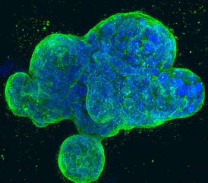
This is a three-dimensional culture of human breast cancer cells, with DNA stained blue and a protein in the cell surface membrane stained green. Credit: National Institutes of Health

Adenosine deaminases are a family of three enzymes encoded by the ADAR genes, which stand for adenosine deaminase acting on RNA. They regulate gene expression by modifying nucleotides within double stranded RNA molecules, serving as fundamental editors in the development of new stem cells.

The enzyme, however, is also activated in cancers as diverse as liver, breast and leukemia. A research team led by senior author Catriona Jamieson, MD, PhD, deputy director of the Sanford Stem Cell Clinical Center and deputy director of the UC San Diego Moores Cancer Center, found that the normal functions of the ADAR1 enzyme are hijacked by pre-malignant cells, leading to a cascade of molecular consequences that promote malignant transformation, dormant cancer stem cell generation and resistance to treatment.
"We were able to illuminate the abilities of ADAR1 to 'hyper-mutate' tumor suppressor RNAs in leukemia and, at the same time, edit the microRNA aimed at targeting the tumor suppressor RNA. This enzyme turns on cancer resistance via a domino effect on RNA instead of DNA," said Qingfei Jiang, PhD, Assistant Project Scientist, Jamieson's lab.
Jamieson characterized RNA editing as tweaking basic genetic blueprints, not fundamentally rewriting them. Nonetheless, the results might be dramatic. "One result of detection of malignant RNA editing could be exposing dormant cancer stem cells that often escape therapies that target dividing cells, which leads to therapeutic resistance and disease relapse, and also highlight ADAR as a potentially tractable target for cancer stem cell elimination," said Jamieson.
Researchers from different part of the world are invited to submit abstract on their unpublished latest research at our upcoming conference Cell Tissue Science 2019 which is focused on the complications and consequences of Stem Cell, Regenerative Medicine, Stem Cell Therapy, Cancer Cell Biology,Technical Advancements in cancer treatment and many more. We as committee members of the conference welcome you to be a part of the conference “ 12th World Congress on Cell & Tissue Science” in Singapore on March 11-12, 2019. 
You can submit your abstract on Session or Track : 09. Stem Cells Biology With Regards to Christmas and New Year Celebration we are providing a special discount of 30% on all Registration Categories for more information please  visit by Click Here

Wednesday, 26 December 2018

Human blood cells can be directly reprogrammed into neural stem cells


Scientists from the German Cancer Research Center (DKFZ) and the stem cell institute HI-STEM* in Heidelberg have succeeded for the first time in directly reprogramming human blood cells into a previously unknown type of neural stem cell. These induced stem cells are similar to those that occur during the early embryonic development of the central nervous system. They can be modified and multiplied indefinitely in the culture dish and can represent an important basis for the development of regenerative therapies.

Stem cells are considered to be the all-rounders of our tissues: they can multiply indefinitely and then if they are pluripotent embryonic stem cells that can generate all conceivable cell types. In 2006, the Japanese scientist Shinya Yamanaka recognized that such cells could also be produced in the laboratory from mature body cells. Four genetic factors alone are sufficient to reverse the course of development and produce so-called induced pluripotent stem cells (iPS) that have identical properties to embryonic stem cells.
"This was a major breakthrough for stem cell research," said Andreas Trumpp, German Cancer Research Center (DKFZ) and Director of HI-STEM in Heidelberg."This applies in particular to for research in Germany, where the generation of human embryonic stem cells is not permitted. Stem cells have enormous potential both for basic research and for the development of regenerative therapies that aim to restore diseased tissue in patients. However, reprogramming is also associated with problems: For example, pluripotent cells can form germ line tumors, so-called teratomas.
Another possibility is not to completely turn back the course of development. For the first time, Trumpp's team has succeeded in reprogramming mature human cells in such a way that a defined type of induced neural stem cells is produced that can multiply almost indefinitely.
"We used four genetic factors like Yamanaka, but different ones for our reprogramming," explains Marc Christian Thier, first author of the study. "We assumed that our factors would allow reprogramming to an early stage of development of the nervous system."
In the past, other research groups also reprogrammed connective tissue cells into mature nerve cells or neural precursor cells. However, these artificially produced nerve cells often could not be expanded and could therefore hardly be used for therapeutic purposes. "Often, it was a heterogeneous mixture of different cell types that might not exist in the body under physiological conditions," said Andreas Trumpp explaining the problems.

Together with stem cell researcher Frank Edenhofer from the University of Innsbruck and neuroscientist Hannah Monyer from DKFZ and the Heidelberg University Hospital, Trumpp and his team have succeeded in reprogramming different human cells: connective tissue cells of the skin or pancreas as well as peripheral blood cells."The origin of the cells had no influence on the properties of the stem cells," said Thier. In particular, the possibility of extracting neural stem cells from the blood of patients without invasive intervention is a decisive advantage for future therapeutic approaches.

What is special about the reprogrammed cells of the Heidelberg researchers is that they are a homogeneous cell type that resembles a stage of neural stem cells that occurs during the embryonic development of the nervous system. "Corresponding cells exist in mice and probably also in humans during early embryonic brain development," said Thier. "We have described here a new neural stem cell type in the mammalian embryo.

These so called "induced Neural Plate Border Stem Cells" (iNBSCs) have a broad development potential. The iNBSCs of the Heidelberg scientists are expandable and multipotent and can develop in two different directions. On the one hand, they can take the path of development to mature nerve cells and their supplier cells, the glial cells, i.e. become cells of the central nervous system. On the other hand, they can also develop into cells of the neural crest, from which different cell types emerge, for example peripheral sensitive nerve cells or cartilage and bones of the skull.

The iNBSCs thus form an ideal basis for generating a broad range of different cell types for an individual patient. "These cells have the same genetic material as the donor and are therefore presumably recognized as "self" by the immune system and are not rejected," explains Thier.
The CRISPR/Cas9 gene scissors can be used to modify the iNBSC or repair genetic defects, as the scientists have shown in their experiments. "They are therefore of interesting both for basic research and the search for new active substances and for the development of regenerative therapies, for example in patients with diseases of the nervous system. However until we can use them in patients, a lot of research work will still be necessary," emphasizes Trumpp.
We welcome researchers from different part of the to submit abstract on their latest research at our upcoming conference Cell Tissue Science 2019 which is mainly focuses on the complications the consequences of Stem Cell, Regenerative Medicine, Stem Cell Therapy, Cancer Cell Biology,Technical Advancements in cancer treatmentand many more.We welcome you to the our upcoming conference “ 12th World Congress on Cell & Tissue Science” . 
You can submit your abstract on Session or Track : 09. Stem Cells Biology

Tuesday, 18 December 2018

Depression, anxiety may take same toll on health as smoking and obesity


An annual physical typically involves a weight check and questions about unhealthy habits like smoking, but a new study from UC San Francisco suggests health care providers may be overlooking a critical question: Are you depressed or anxious?

Anxiety and depression may be leading predictors of conditions ranging from heart disease and high blood pressure to arthritis, headaches, back pain and stomach upset, having similar effects as long-established risk factors like smoking and obesity, according to the new research.
In the study looked at the health data of more than 15,000 older adults over a four-year period.
They found that 16 percent (2,225) suffered from high levels of anxiety and depression, 31 percent (4,737) were obese and 14 percent (2,125) were current smokers, according to their study published in the journal Health Psychology on Dec. 17, 2018.

Participants with high levels of anxiety and depression were found to face 65 percent increased odds for a heart condition, 64 percent for stroke, 50 percent for high blood pressure and 87 for arthritis, compared to those without anxiety and depression.
"These increased odds are similar to those of participants who are smokers or are obese," said O'Donovan, who, with Niles, also is affiliated with UCSF Weill Institute for Neurosciences. "However, for arthritis, high anxiety and depression seem to confer higher risks than smoking and obesity."
Cancer an Exception to Conditions Impacted by Depression and Anxiety
Unlike the other conditions investigated, the authors found that high levels of depression and anxiety were not associated with cancer incidence. This confirms results from previous studies, but contradicts a prevailing idea shared by many patients.
"Our findings are in line with a lot of other studies showing that psychological distress is not a strong predictor of many types of cancer," O'Donovan said. "On top of highlighting that mental health matters for a whole host of medical illnesses, it is important that we promote these null findings. We need to stop attributing cancer diagnoses to histories of stress, depression and anxiety."
Niles and O'Donovan discovered that symptoms such as headache, stomach upset, back pain and shortness of breath increased exponentially in association with high stress and depression. Odds for headache, for example, were 161 percent higher in this group, compared with no increase among the participants who were obese and smokers.
Treating Mental Health Can Cut Health Care Costs
"Anxiety and depression symptoms are strongly linked to poor physical health, yet these conditions continue to receive limited attention in primary care settings, compared to smoking and obesity," Niles said. "To our knowledge this is the first study that directly compared anxiety and depression to obesity and smoking as prospective risk factors for disease onset in long-term studies."
The results of the study underscore the "long-term costs of untreated depression and anxiety," said O'Donovan. "They serve as a reminder that treating mental health conditions can save money for health systems."
The two authors evaluated health data from a government study of 15,418 retirees, whose average age was 68. Depression and anxiety symptoms were assessed using data from participant interviews. Participants were questioned about their current smoking status, while weight was self-reported or measured during in-person visits. Medical diagnoses and somatic symptoms were reported by participants.
We welcome researchers from different part of the to submit abstract on their latest research at our upcoming conference Cell Tissue Science 2019 which is mainly focuses on the complications the consequences of Stem Cell, Regenerative Medicine, Stem Cell Therapy, Cancer Cell Biology , Technical Advancements in cancer treatment and many more.We welcome you to the our upcoming conference “ 12th World Congress on Cell & Tissue Science” . 
You can submit your abstract on Session or Track : 08.Cancer Cell Biology

Saturday, 15 December 2018

The source of stem cells points to two proteins

New MSU research published in the journal eLife has pinpointed two proteins that are the keys to stem cell creation.
Credit: Courtesy of MSU
Mammalian embryos are unlike those of any other organism as they must grow within the mother's body. While other animal embryos grow outside the mother, their embryonic cells can get right to work accepting assignments, such as head, tail or vital organ. By contrast, mammalian embryos must first choose between forming the placenta or creating the baby.

New research at Michigan State University and published in the journal eLife has pinpointed two proteins that are the keys to this decision making. The process of assigning cells to placenta or baby is important because that is when pluripotent cells are made. These adaptable pluripotent cells are critical to stem cell research, and these two proteins could be the key to deciphering how pluripotent cells are born, said Amy Ralston, MSU's inaugural James K. Billman Jr., M.D. endowed professor and the study's senior author.
"Pluripotent cells are the progenitors of embryonic stem cells, and they are famous because they can become any part of the body," she said. "We have discovered a process that regulates the balance between pluripotent and placenta, and it works by changing the physical location of cells within the embryo's ball of cells."
To form this wondrous ball, the mother packages two closely related proteins into her eggs, which help oversee this decision-making process. The scientific team used genetic tools to eliminate the proteins YAP1 and WWTR1 from mouse eggs and embryos. The team discovered that these two proteins first position these cells into distinct inside and outside locations, which then decides their fate: placenta or pluripotent.

Each phase of Ralston's research peels back a layer into the creation of stem cells, a process that nature performs with 100 percent efficiency. On the other end of the spectrum, lab-created stem cells are created with 1 percent efficiency.
"Obviously, our understanding of how nature creates stem cells is incomplete," said Tristan Frum, MSU biochemist and molecular biologist"We've suspected these proteins were involved in creating stem cells, but our work reveals that they do so in a surprising way."
Cells of the early embryo express proteins that make them "stick" to the outside of the embryo.
"We've identified a way that cells evade this stickiness, crawl inside the embryo and acquire the properties that make stem cells so interesting and useful," Frum said. "When we make stem cells in the lab, they must acquire a unique cell membrane. Our work shows how nature does this and provides clues that can guide us to control stem cells more efficiently for use in medicine".
For example, the team's discoveries could lead to advances in stem cell technologies using organoids, which are stem cell-derived mini organs. Organoidsare an exciting new paradigm in stem cell and regenerative medicine, The similarities between embryos and organoids are remarkable. Therefore, by studying how the mouse embryo builds itself, we may one day build organs from human stem cells." says Ralston.
We welcome researchers from different part of the to submit abstract on their latest research at our upcoming conference Cell Tissue Science 2019 which is mainly focuses on the complications the consequences of Stem Cell, Regenerative Medicine, Stem Cell Therapy, Cancer Cell Biology,Technical Advancements in cancer treatmentand many more.We welcome you to the our upcoming conference “ 12th World Congress on Cell & Tissue Science” . 
You can submit your abstract on Session or Track : 09. Stem Cells and its Applications

Tuesday, 11 December 2018

Potential seen for tailoring treatment for acute myeloid leukemia


Advances in rapid screening of leukemia cells for drug susceptibility and resistance are bringing scientists closer to patient-tailored treatment for acute myeloid leukemia (AML).

Research on the drug responses of leukemia stem cells may reveal why some attempts to treat are not successful or why initially promising treatment results are not sustained.

AML is a serious disorder of certain blood-forming cells. In this disease, certain early precursor cells in the bone marrow that usually develop into white blood cells don't mature properly. They remain frozen as primitive cells called blasts, unable to further differentiate and mature. These can accumulate and cause low blood counts that reduce the ability to fight infections, and low platelet counts that cause risk of life threatening hemorrhage.

Leukemia stem cells -- the progenitors for the immature, cancerous blood cells -- propagate AML, and also play a role in the cancer returning after treatment. Cancer researchers are interested in how genes are expressed in this cell population, because this data may hold clues to resistance to standard therapies and answers to why some patients relapse.

A study presented at the 60th Annual Meeting of the American Society of Hematology in San Diego looked at the drug response patterns of stem cells and blast cells taken from individual patients diagnosed with acute myeloid leukemia. The information was gathered through high throughput screening, a state of the art method for quickly evaluating and testing many samples.

The researchers found that leukemia stem cells and blast cells diverged in their drug susceptibility patterns, and also that these patterns differed from patient to patient.
For example: Blast cells s responded in the test to the drugs most commonly used to treat patients, but none were effective against leukemia stem cells. The researchers did find 12 drugs from eight classes that seemed to preferentially target leukemia stem cells, compared to blast cells. Many of them are not often used in patients with this type of cancer.
The multidisciplinary team on the project included stem cell biologists, hematologists, medical oncologists, pathologists, computer scientists, drug developers and others.

The senior researcher was hematologist Dr. Pamela Becker, professor of medicine at the UW School of Medicine. She is also a scientist at the Fred Hutchinson Cancer Research Center and the UW Medicine Institute for Stem Cells and Regenerative Medicine, and sees patients with blood disorders at the Seattle Cancer Care Alliance.

In the laboratory study, the researchers compared the drug sensitivity of blast cell and stem cell populations taken from the same six patients. In doing so, they tested a custom panel of drugs, targeted agents and drug combinations on the cells, and did genetic analyses for 194 mutations. The panel included both FDA approved and investigational drugs.

The unique drug susceptibility patterns observed in leukemia stem cells and blast cells are leading the scientists to hope that patient-specific approaches could be developed against acute myeloid leukemia, with the goal of improving the outcomes for people with this form of blood cancer.
We welcome researchers from different part of the to submit abstract on their latest research at our upcoming conference Cell Tissue Science 2019 which is mainly focuses on the complications the consequences of Stem Cell, Regenerative Medicine, Stem Cell Therapy, Cancer Cell Biology , Technical Advancements in cancer treatmentand many more.We welcome you to the our upcoming conference “ 12th World Congress on Cell & Tissue Science” . 
You can submit your abstract on Session or Track : 09. Stem Cells and its Applications

Thursday, 6 December 2018

Citrate-based biomaterial fuels bone healing with less rejection


A material based on a natural product of bones and citrus fruit, called citrate, provides the extra energy that stem cells need to form new bone tissue, according to a team of Penn State bioengineers. Their new understanding of the mechanism that allows citrate to aid in bone regeneration will help the researchers develop slow-release, biodegradable, citrate-releasing scaffolds to act as bone-growth templates to speed up healing in the body.
"In our lab, we have been working with citrate for over a decade," said Jian Yang, Professor of Biomedical Engineering, Penn State. "We knew that in the human body, 90 percent of organic citrate is located in skeletal tissue. But no one had really tried to use citrate as a building block to make bone biomaterials. Our new paper tries to understand how citrate helps in bone healing and uses the understanding to guide the design of new biomimetic biomaterials for better bone repair."
Autografting -- taking bone from another part of the patient's body and grafting it to the wound -- is the method used most for bone regeneration in a hospital setting. This is not always a suitable method, especially in the case of large wounds or bone tissue removed during cancer treatment.

Synthetic biomaterials would be a welcome replacement and many labs are working on developing them. But current synthetic materials cause significant inflammation, and the bone healing rate is slow and healing quality is poor. The body encapsulates the implant with fibrotic tissues that keep the implant from integrating with surrounding bone. With Yang's material, the researchers do not see encapsulation, and chronic inflammation is minimal.

Chuying Ma, a doctoral student in Yang's lab, is lead author on the paper. Ma was given the problem of uncovering the poorly known mechanism underlying the body's use of citrate to regenerate bone. She found that the stem cell membrane has a transporter that is used to transport citrate into the cell to elevate the cellular energy level. When the bone stem cells differentiate to make new bone cells, they require more energy as support for active bone formation. The timing and dosage of citrate supply to stem cells are also critical. Yang and Ma coined the newly identified citrate effect on stem cell differentiation as "metabonegenic regulation" in the paper.

The team also identified a second factor involved in energy production, an amino acid called phosphoserine. With their new understanding of the mechanism for bone regrowth, they developed a biomaterial incorporating both citrate and phosphoserine and tested it on rat models.
"Using our new material, we see the early deposition of new bone at one month," Ma said. "This is much earlier than the biomaterials widely used in FDA-approved devices. In this study we tested two models, the femoral condyle bone and cranial bone defects."
In both animal models we see the new biomaterial is better than the commercial materials in inducing early bone formation and also promoting bone maturation, according to Ma. "To me, this is an important finding," said Yang, who is a faculty member in Penn State's Materials Research Institute and the Huck Institutes of the Life Sciences. "Citrate is now recognized as a central linker between stem cell metabolism and differentiation. We are uncovering the mechanism whereby citrate influences stem cell activity, not only in bone, but by implication extending to other types of cells and tissues. For instance, there is a high concentration of citrate in the cerebrospinal fluid surrounding the brain. People can now use this understanding to start looking at citrate as a metabolism regulator to further regulate stem cells for other types of tissues and organs throughout the body."
We welcome researchers from different part of the to submit abstract on their latest research at our upcoming conference Cell Tissue Science 2019 which is mainly focuses on the complications the consequences of Stem Cell, Regenerative Medicine, Stem Cell Therapy, Cancer Cell Biology , Technical Advancements in cancer treatment and many more.We welcome you to the our upcoming conference “ 12th World Congress on Cell & Tissue Science” . 
You can submit your abstract on Session or Track : 10.Biomaterial and Bioengineering

Tuesday, 4 December 2018

Healthy blood stem cells have as many DNA mutations as leukemic cells


Researchers from the Princess Máxima Center for Pediatric Oncology have shown that the number of mutations in healthy and leukemic blood stem cells does not differ. Rather the location of the mutations in the DNA is relevant. Using the mutation patterns in the hematopoietic stem and progenitor cells (HSPCs) the team was able to trace the developmental lineage tree of the cells.

Mutational load
"Blood stem cells divide about once every 40 weeks," says Van Boxtel, "and we saw that eleven mutations occur during one division." The older the test subject, the more mutations the researchers found because the mutations accumulate over the years. Yet, these people were fit as a fiddle.Nevertheless, mutations in blood stem cells may also lead to leukemia. "We thought that people with leukemia would have more mutations than healthy people," says Van Boxtel, "but this is not the case." The HSPCs of patients with acute myeloid leukemia (AML) contain as many mutations as those from healthy people. The researchers published their results in the open access journal Cell Reports.
Developmental lineage tree
The researchers also describe how they were able to trace the developmental lineage tree of hematopoiesis using the mutation pattern of HSPCs. "If you study the pattern of mutations of a cancer cell, you can figure out which cell it comes from," explains Van Boxtel. "We have shown this for HSPCs now, but especially for solid tumors the origin of the cancer cell is very relevant for selecting the most effective treatment strategy." The technique has a lot of potential according to the authors. The next step for Van Boxtel and his team will be to study the origin of causative mutations in second cancers in survivors of pediatric cancer.
"So far, we assumed that new mutations occur as a result of intensive treatment during childhood and cause second cancers later in life. We can now test whether these mutations are indeed new or already existed and contributed to both incidences of cancer. This is relevant knowledge when making a treatment plan for children with cancer."
We welcome researchers from different part of the to submit abstract on their latest research at our upcoming conference Cell Tissue Science 2019 which is mainly focuses on the complications the consequences of Stem Cell, Regenerative Medicine, Stem Cell Therapy, Cancer Cell Biology , Technical Advancements in cancer treatment and many more.We welcome you to the our upcoming conference “ 12th World Congress on Cell & Tissue Science” . 
You can submit your abstract on Session or Track : 9.Stem Cells and its Applications

Tuesday, 6 November 2018

Advance stem cell therapy with biodegradable scaffold


181105081710_1_540x360
A biodegradable inorganic nano-scaffold, consisting of stem cells, proteins and drugs, for advanced stem cell therapy and drug delivery.
Credit: KiBum Lee, Letao Yang and Sy-Tsong Dean Chueng

Stem cell transplantation, which shows promise as a treatment for central nervous system diseases, has been hampered by low cell survival rates, incomplete differentiation of cells and limited growth of neural connections.

So, Rutgers scientists designed bio-scaffolds that mimic natural tissue and got good results in test tubes and mice, according to a study in Nature Communications. These nano-size scaffolds hold promise for advanced stem cell transplantation and neural tissue engineering. Stem cell therapy leads to stem cells becoming neurons and can restore neural circuits.
"It's been a major challenge to develop a reliable therapeutic method for treating central nervous system diseases and injuries," said study senior author KiBum Lee, Department of Chemistry and Chemical Biology at Rutgers University-New Brunswick. "Our enhanced stem cell transplantation approach is an innovative potential solution."
The researchers, in cooperation with neuroscientists and clinicians, plan to test the nano-scaffolds in larger animals and eventually move to clinical trials for treating spinal cord injury. The scaffold-based technology also shows promise for regenerative medicine.
We welcome researchers from different part of the world to submit your latest research at our upcoming conference “12th World Congress on Cell & Tissue Science” scheduled on March 11-12,2019 in Singapore which is mainly focuses on the complications the consequences of Stem Cell, Regenerative Medicine, Stem Cell Therapy, Cancer Cell Biology , Technical Advancements in cancer treatment and many more. For more info visit our conference website:Cell Tissue Science 2019

Monday, 5 November 2018

Novel method to block immunosuppression in cancer


Belgian research groups from the UCLouvain and WELBIO, VIB and Ghent University, and the biotechnology company Aargenx elucidated the three-dimensional structure of an assembly of proteins operating on cells that dampen immune responses. They also discovered how an antibody can block this assembly and the immunosuppression it induces downstream. Such an antibody could serve to stimulate immunity against tumor cells in cancer patients, triggering the destruction of their tumors by immune cells. The study is published in the authoritative Journal Science.

Immunosuppression through a cascade of interactions

Tregs (regulatory T lymphocytes) are immunosuppressive cells that normally counterbalance excessive immune reactions to prevent autoimmune diseases. But in cancer patients, they play deleterious roles by tempering immune reactions against tumor cells. Tregs induce their effects by producing a protein messenger called TGF-beta. This messenger transmits inhibitory signals to immune cells in the immediate vicinity, notably those that are supposed to destroy tumors in cancer patients.

The way Tregs produce TGF-beta is complex and finely regulated, because TGF-beta is very potent and must be kept under tight control. Three years ago, Prof. Sophie Lucas and her team at the de Duve Institute of the UCLouvain discovered that TGF-beta is released by Tregs from a protein called GARP, present on the Treg surface.

In collaboration with argenx, her team also discovered that it was possible to block the release of TGF-beta from GARP with specific antibodies, which were rare and difficult to obtain. The next thing to find out was how GARP regulates the production of the TGF-beta messenger and how antibodies actually block its release.

The molecular mechanisms elucidated

Sophie Lucas and argenx initiated a collaboration with the team of Prof. Savvas Savvides at the VIB-UGent Center for Inflammation Research, to resolve the three-dimensional structure of the protein assembly made of GARP and TGF-beta.

The researchers used X-ray crystallography, a method that has been used to study the structure of molecules for more than a century and that is still being developed for the study of biological macromolecules at atomic resolution. However, they were confronted with the practical problem that they could not readily obtain crystals of the GARP and TGF-beta complex.

Via a highly collaborative effort spearheaded by Dr. Stéphanie Lienart (UCLouvain) and Dr. Romain Merceron (VIB -- Ghent University), the two teams decided to use a blocking antibody to stabilize the structure -- a successful approach that not only helped to generate suitable crystals for structure determination, but also provided details about how a therapeutic antibody might function.
Prof. Savvas Savvides (VIB-UGent): "We discovered that GARP resembles a horseshoe that is straddled by TGF-beta. The two molecules are so intricately assembled that TGF-beta itself contributes to the structure of the GARP horseshoe. The antibody fragment sticks to both GARP and TGF-beta in the assembly. It appears to glue the two molecules to one another, ensuring that when other molecules pull on one part of the assembly, the small, active part of TGF-beta is not released, and is thus prevented from conveying its inhibitory message." 
Prof. Sophie Lucas (de Duve Institute at the UCLouvain): "Visualization of this large molecular assembly illustrates the feasibility of blocking TGF-beta activity emanating from a precisely defined and restricted cellular source, such as the surface of Tregs. This can lead to the design of exquisitely specific approaches to treat various diseases associated with altered TGF-beta or Treg activity, most notably for the immunotherapy of cancer." 
We welcome researchers from different part of the world to submit your latest research at our upcoming conference “12th World Congress on Cell & Tissue Science” scheduled on March 11-12,2019 in Singapore which is mainly focuses on the complications the consequences of Stem Cell, Regenerative Medicine, Stem Cell Therapy, Cancer Cell Biology , Technical Advancements in cancer treatment and many more. For more info visit our conference website:Cell Tissue Science 2019

Saturday, 3 November 2018

Normal function of ALS and dementia linked gene determined for the first time


The normal function of a gene associated with the neurodegenerative diseases amyotrophic lateral sclerosis (ALS) and frontotemporal dementia (FTD) has been determined for the first time by University of Bath scientists.

Both ALS, the most common form of motor neurone disease, and FTD progressively develop in adults and have been shown to be closely linked, with overlapping genetic origins and similar effects on some nervous system cells. There is no cure for either disease.

One gene, called C9orf72, is strongly suspected to be the cause of inherited ALS and FTD when a short sequence of DNA repeats hundreds or even thousands of times within the gene, causing problems in its transcription and expression. However, until now scientists were uncertain as to what normal role or roles the C9orf72 gene played in cells.

A University of Bath team from the Department of Biology and Biochemistry have now biochemically characterised the protein produced by the C9orf72 gene and have shown that it is a guanine nucleotide exchange factor (GEF).

GEFs are a type of protein which regulate molecules called RABs which control key processes inside cells. They respond to signals received by a cell in normal as well as diseased states. Recent evidence suggests that some GEF proteins may be potential therapeutic targets for developing drugs to treat various diseases, including cancer.
Dr Vasanta Subramanian said: "It is vital that we know as much as we can about the causes and triggers for ALS and FTD, so we have a better chance of developing treatments, and even one day a cure for these devastating diseases.Understanding the function of C9orf72, which is linked to both diseases, is a crucial step in that process."
 "These findings are very timely, as new gene therapy approaches are being developed to specifically target the C9orf72 gene. Understanding what the healthy gene is doing within cells will be important in ensuring that the toxic and not the beneficial effects of the gene are targeted"said Dr Brian Dickie, Director of Research, Motor Neurone Disease Association 
We welcome researchers from different part of the world to submit your latest research at our upcoming conference “12th World Congress on Cell & Tissue Science” scheduled on March 11-12,2019 in Singapore which is mainly focuses on the complications the consequences of Stem Cell, Regenerative Medicine, Stem Cell Therapy, Cancer Cell Biology , Technical Advancements in cancer treatment and many more. For more info visit our conference website:Cell Tissue Science 2019

Friday, 2 November 2018

New epigenetic drug strategy to treat cancer


Researchers have discovered that inhibiting CDK9, a DNA transcription regulator, reactivates genes that have been epigenetically silenced by cancer. Reactivation leads to restored tumor suppressor gene expression and enhanced anti-cancer immunity. It is the first time this particular kinase has been linked to gene silencing in mammals.

It has been established that epigenetic mediators of gene silencing present new targets for cancer drugs. Hanghang Zhang, PhD, Fels Institute for Cancer Research & Molecular Biology, Lewis Katz School of Medicine at Temple University (LKSOM), performed a live cell drug screen with genetic confirmation to identify CDK9 as a target and to develop and test an effective inhibitor -- MC180295.

The new drug is highly selective, potentially avoiding the side effects associated with inhibiting the cell cycle. In the study it showed broad effectiveness against cancer both in vitro and in vivo. The drug was discovered in collaboration with investigators at the Moulder Center for Drug Discovery at the Temple University School of Pharmacy.
"In addition to reactivating tumor suppressor genes, CDK9 inhibition induces sensitivity to the immune checkpoint inhibitor ?-PD-1 in vivo," said Issa. "It is an excellent target for epigenetic cancer therapy."
 
We welcome researchers from different part of the world to submit your latest research at our upcoming conference “12th World Congress on Cell & Tissue Science” scheduled on March 11-12,2019 in Singapore which is mainly focuses on the complications the consequences of Stem Cell, Regenerative Medicine, Stem Cell Therapy, Cancer Cell Biology , Technical Advancements in cancer treatment and many more. For more info visit our conference website:Cell Tissue Science 2019

Saturday, 29 September 2018

Enzymes 'partner up' to accelerate cancer, aging diseases

A new study from molecular biologists at Indiana University has identified cellular processes that appear to supercharge both the growth and shrinkage of the chemical "caps" on chromosomes associated with aging, called telomeres.

The work, focused on two enzymes in yeast, could lead to new insights on stopping runaway cellular growth in cancer tumors, as well as the treatment of premature aging disorders such as progeria (aka "Benjamin Button disease").
"This work confirms that two specific enzymes are called helicases which are involved in telomere maintenance, and demonstrates they're even stronger in combination.This is significant since dysfunction in telomere maintenance has been found in 100 percent of cancers. Literally, 100 percent. So, it's very likely they play a role in the disease."said Matthew Bochman, Associate Professor, IU Bloomington College of Arts and Sciences 
Helicases are enzymes that unwind double-stranded DNA into a single stand for the purposes of replication, recombination and repair. In humans, the RecQ4 helicase functions similarly to the Hrq1 helicase in yeast. The Pif1 helicase is the same in both species.

In healthy people, telomeres shorten slowly over the lifespan as part of the natural aging process. In cancer cells, the brakes come off this process, so telomeres never grow shorter -- resulting in uncontrolled cellular replication. In people with premature aging disorders, telomeres rapidly shrink, resulting in death from "old age" in the late teenage years.

Mutations of the Pif1 helicase have been linked to several types of cancer, including common forms such as breast, ovarian and colon cancer. Mutations in the RecQ4 helicase have been linked to three different diseases associated with predispositions for cancer.

The study specifically found that Hrq1 and Pif1 are a "dynamic duo" that combine to create a "super inhibitor" or, under certain specific conditions, a "super stimulator" of telomere growth.
"Now, we can now really start to pick apart more about how these enzymes work together or alone in cancer cells," Bochman said.
The work may help scientists better understand whether certain cancers involve errors in DNA recombination, DNA repair or telomere maintenance or some other mechanistic problem. This, in turn, could lead to new ways to disrupt or harness these processes with drugs or other therapies.
 
We welcome researchers from different part of the to submit abstract on their latest research at our upcoming conference Cell Tissue Science 2019 which is mainly focuses on the complications the consequences of Stem Cell, Regenerative Medicine, Stem Cell Therapy, Cancer Cell Biology , Technical Advancements in cancer treatment and many more.We welcome you to the our upcoming conference “ 12th World Congress on Cell & Tissue Science” . For more info visit :Cell Tissue Science 2019