Showing posts with label Cell Tissue Science 2019. Show all posts

Saturday, 15 June 2019

'Virtual biopsy' device to detect skin tumors


Using sound vibrations and pulses of near-infrared light, a Rutgers University scientist has developed a new "virtual biopsy" device that can quickly determine a skin lesion's depth and potential malignancy without using a scalpel.

The ability to analyze a skin tumor non-invasively could make biopsies much less risky and distressing to patients, according to a report in Wiley Online Library. Currently, physicians who perform surgical biopsies often don't know the extent of a lesion and whether it will be necessary to refer the patient to a specialist for extensive tissue removal or plastic surgery until surgery has already begun.

The first-of-its-kind experimental procedure, called vibrational optical coherence tomography (VOCT), creates a 3-D map of the legion's width and depth under the skin with a tiny laser diode. It also uses soundwaves to test the lesion's density and stiffness since cancer cells are stiffer than healthy cells. An inch-long speaker applies audible soundwaves against the skin to measure the skin's vibrations and determine whether the lesion is malignant.
"This procedure can be completed in 15 minutes with no discomfort to the patient, who feels no sensation from the light or the nearly inaudible sound. It's a significant improvement over surgical biopsies, which are invasive, expensive and time consuming," said Frederick Silver, a professor of pathology and laboratory at Rutgers Robert Wood Johnson Medical School.
The study found that a prototype VOCT device, which awaits FDA approval for large-scale testing, is able to accurately distinguish between healthy skin and different types of skin lesions and carcinomas. The researchers tested the device over six months on four skin excisions and on eight volunteers without skin lesions. Further studies are needed to fine-tune the device's ability to identify a lesion's borders and areas of greatest density and stiffness, which would allow physicians to remove tumors with minimally invasive surgery.

An important announcement regarding our upcoming conference 12th World Congress on Cell & Tissue Science (Cell Tissue Science 2019) scheduled on September 13-14,2019 in Singapore. You can also present your latest research at the different topics such as Cancer Cell BiologyStem Cell & its applications and many more along with other distinguished professors, doctors and researchers from all over the world.
If interested kindly proceed with submitting your abstract and latest biography along with a photography to our online abstract submission page given below: Link for submission: Click Here
Source: https://www.sciencedaily.com/releases/2019/06/190613103129.htm

Friday, 7 June 2019

New genes out of nothing


One key question in evolutionary biology is how novel genes arise and develop. Swedish researchers now show how new genes and functions that are advantageous to bacteria can be selected from random DNA sequences. The results are presented in the scientific journal mBio.

How do new genes and functional proteins arise and develop? This is one of the most fundamental issues in evolutionary biology. Two different types of mechanism have been proposed: (1) new genes with novel functions arise from existing genes, and (2) new genes and proteins evolve from random DNA sequences with no similarity to existing genes and proteins.

In the present study, the researchers explored the latter type of mechanism: evolution of new genes and proteins from randomised DNA sequences as its called as de novo evolution. It is fairly easy to understand that when a gene already exists, it can be modified and acquire a new function. But how does "nothing" turn into a function affording a small advantage that is favoured by natural selection?

The raw material for the experiment was an big library of some 500 million randomised gene sequences, from which peptide sequences with a biological function were identified. In the experiment, random gene sequences were placed on a plasmid and overexpressed. The scientists then investigated whether they could give bacteria a specific, defined property. Were they, for example, able to give the bacteria antibiotic resistance? They identified several short peptides (22-25 amino acids long) that could give the bacteria a high degree of resistance to aminoglycosides, an important class of antibiotics used for severe infections.
"When the project started, we had low expectations. We were amazed when we found peptides able to confer a resistance level 48 times higher," says Dr Michael Knopp, the study's lead author.
Through a combination of genetic and functional experiments, the scientists were able to demonstrate that the peptides cause resistance by attaching themselves to bacterial cell membranes and affecting the proton potential across the membrane. The disruption of the proton potential causes a decrease in antibiotic uptake, rendering the bacteria resistant.
"This study is important because it shows that completely random sequences of amino acids can give rise to new, advantageous functions, and that this process of de novo evolution can be studied experimentally in the laboratory," says Dan I. Andersson, Professor of Medical Bacteriology, who is chiefly responsible for the study.
An important announcement regarding our upcoming conference 12th World Congress on Cell & Tissue Science (Cell Tissue Science 2019) scheduled on September 13-14,2019 in Singapore. You can also present your latest research at the different topics such as Cancer Cell BiologyStem Cell & its applications and many more along with other distinguished professors, doctors and researchers from all over the world.
If interested kindly proceed with submitting your abstract and latest biography along with a photography to our online abstract submission page given below: Link for submission: Click Here
Source of the article: https://www.sciencedaily.com/releases/2019/06/190604131214.htm

Saturday, 1 June 2019

How prostate cancer cells mimic bone when they metastasize


Prostate cancer often becomes lethal as it spreads to the bones, and the process behind this deadly feature could potentially be turned against it as a target for bone-targeting radiation and potential new therapies.

Study published online in the journal PLOS ONE, Duke Cancer Institute researchers describe how prostate cancer cells develop the ability to mimic bone-forming cells called osteoblasts, enabling them to proliferate in the bone microenvironment.

Attacking these cells with radium-233, a radioactive isotope that selectively targets cells in these bone metastases, has been shown to prolong patients' lives. But a better understanding of how radium works in the bone was needed.

The mapping of this mimicking process could lead to a more effective use of radium-233 and to the development of new therapies to treat or prevent the spread of prostate cancer to bone.
"Given that most men who die of prostate cancer have bone metastases, this work is critical to helping understand this process," said lead author Andrew Armstrong, Director of Research at the Duke Cancer Institute Center for Prostate and Urologic Cancers.
The research team enrolled a small study group of 20 men with symptomatic bone-metastatic prostate cancer. When analyzing the circulating tumor cells from study participants, they found that bone-forming enzymes appeared to be expressed commonly, and that genetic alterations in bone forming pathways were also common in these prostate cancer cells.

They validated these new genetic findings in a separate multicenter trial involving a larger group of more than 40 men with prostate cancer and bone metastases.

Following treatment with radium-223, the researchers found that the radioactive isotope was concentrated in bone metastases, but tumor cells still circulated and cancer progressed within six months of therapy. The researchers found a range of complex genetic alterations in these tumor cells that likely enabled them to persist and develop resistance to the radiation over time.
"Osteomimicry may contribute in part to how prostate cancer spreads to bone, but also to the uptake of radium-223 within bone metastases and may thereby enhance the therapeutic benefit of this bone targeting radiotherapy," Armstrong said.
He said by mapping this lethal pathway of prostate cancer bone metastasis, the study points to new targets and thus critical areas of research into designing better tumor-targeting therapies.
An important announcement regarding our upcoming conference 12th World Congress on Cell & Tissue Science (Cell Tissue Science 2019) scheduled on September 13-14,2019 in Singapore. You can also present your latest research at the different topics such as Cancer Cell Biology, Stem Cell & its applications and many more along with other distinguished professors, doctors and researchers from all over the world.
If interested kindly proceed with submitting your abstract and latest biography along with a photography to our online abstract submission page given below: Link for submission: Click Here

Thursday, 24 January 2019

Energizing the immune system to eat cancer


Immune cells called macrophages are supposed to serve and protect, but cancer has found ways to put them to sleep. Now researchers at the Abramson Cancer Center of the University of Pennsylvania say they've identified how to fuel macrophages with the energy needed to attack and eat cancer cells. It is well established that macrophages can either support cancer cell growth and spread or hinder it. But most tumors also express a signal called CD47, which can lull macrophages into a deep sleep and prevent them from eating. Researchers have found that rewiring macrophage metabolism can overcome this signal and act like an alarm clock to rouse and prepare macrophages to go to work. Their findings were published in Nature Immunology today.

Macrophages are immune cells just like T and B cells, but differ in that they can eat cells that are not supposed to be in the body. In fact, they are the most prominent immune cell found in cancer, but unfortunately, most are often convinced to help cancer grow and spread. Cancer cells frequently stop macrophages from attacking them by expressing CD47, a "don't eat me" signal. Researchers now say that merely blocking inhibitory signals like CD47 is not always sufficient to convince macrophages to attack cancer. Instead, two signals are required. First, they need a signal to activate them -- such as a toll-like receptor agonist. After that, a second signal -- such as a CD47 inhibitor -- can lower the threshold needed to wage battle on the cancer.

"It turns out macrophages need to be primed before they can go to work, which explains why solid tumors may resist treatment with CD47 inhibitors alone," said the study's senior author Gregory L. Beatty, Assistant Professor of Hematology-Oncology at Penn's Perelman School of Medicine. 
The team used this approach by activating macrophages with CpG, a toll-like receptor agonist that sends the first signal, and found that it rapidly induced shrinkage of tumors and prolonged survival of mice even without the requirement of T cells. Unexpectedly, they also found that the activated macrophages were able to eat cancer cells even in the presence of high levels of CD47.

To understand the molecular basis of this phenomenon, the team traced the metabolic activity of macrophages and determined that activated macrophages began to utilize both glutamine and glucose as fuel to support the energy requirements needed for them to eat cancer cells. This rewiring of the macrophages metabolism was necessary for CpG to be effective, and the researchers say these findings point to the importance of macrophage metabolism in determining the outcome of an immune response.
"Cancer does not shrink without the help of macrophages and macrophages need the right fuel to eat cancer cells and shrink tumors," Liu said. "To do this, a shift in metabolism is needed to steer the energy in the right direction. It is the metabolism that ultimately allows macrophages to override signals telling them not to do their job."
Beatty points out that patients with diabetes, cardiovascular disease, and other conditions are routinely treated with drugs that could affect macrophage metabolism, but virtually nothing is known about how these drugs might impact immunotherapy responses in cancer, meaning the team's discovery has implications even for existing treatments.
Researchers from different part of the world are invited to submit abstract on their unpublished latest research at our upcoming conference Cell Tissue Science 2019 which is focused on the complications and consequences of Stem CellRegenerative MedicineStem Cell Therapy, Cancer Cell Biology,Technical Advancements in cancer treatment and many more. We as committee members of the conference welcome you to be a part of the conference “ 12th World Congress on Cell & Tissue Science” in Singapore on March 11-12, 2019
You can submit your abstract on Session or Track : 08. Advancement in Cancer Treatments

Wednesday, 23 January 2019

New nanoparticle targets tumor-infiltrating immune cells, flips switch


Immunotherapy's promise in the fight against cancer drew international attention after two scientists won a Nobel Prize this year for unleashing the ability of the immune system to eliminate tumor cells.

But their approach, which keeps cancer cells from shutting off the immune system's powerful T-cells before they can fight tumors, is just one way to use the body's natural defenses against deadly disease. A team of Vanderbilt University bioengineers today announced a major breakthrough in another: penetrating tumor-infiltrating immune cells and flipping on a switch that tells them to start fighting. The team designed a nanoscale particle to do that and found early success using it on human melanoma tissue.
"Tumors are pretty conniving and have evolved many ways to evade detection from our immune system," said John T. Wilson, Assistant Professor of Chemical and Biomolecular Engineering and Biomedical Engineering. "Our goal is to rearm the immune system with the tools it needs to destroy cancer cells.Checkpoint blockade has been a major breakthrough, but despite the huge impact it continues to have, we also know that there are a lot of patients who don't respond to these therapies. We've developed a nanoparticle to find tumors and deliver a specific type of molecule that's produced naturally by our bodies to fight off cancer."
That molecule is called cGAMP, and it's the primary way to switch on what's known as the stimulator of interferon genes (STING) pathway: a natural mechanism the body uses to mount an immune response that can fight viruses or bacteria or clear out malignant cells. Wilson said his team's nanoparticle delivers cGAMP in a way that jump-starts the immune response inside the tumor, resulting in the generation of T-cells that can destroy the tumor from the inside and also improve responses to checkpoint blockade.

While the Vanderbilt team's research focused on melanoma, their work also indicates that this could impact treatment of many cancers, Wilson said, including breast, kidney, head and neck, neuroblastoma, colorectal and lung cancer.

His findings appear today in a paper titled "Endosomolytic Polymersomes Increase the Activity of Cyclic Dinucleotide STING Agonists to Enhance Cancer Immunotherapy" in the journal Nature Nanotechnology.

Daniel Shae, a Ph.D. student on Wilson's team and first author of the manuscript, said the process began with developing the right nanoparticle, built using "smart" polymers that respond to changes in pH that he engineered to enhance the potency of cGAMP. After 20 or so iterations, the team found one that could deliver cGAMP and activate STING efficiently in mouse immune cells, then mouse tumors and eventually human tissue samples.
"That's really exciting because it demonstrates that, one day, this technology may have success in patients," Shae said.
Researchers from different part of the world are invited to submit abstract on their unpublished latest research at our upcoming conference Cell Tissue Science 2019 which is focused on the complications and consequences of Stem CellRegenerative MedicineStem Cell Therapy, Cancer Cell Biology,Technical Advancements in cancer treatment and many more. We as committee members of the conference welcome you to be a part of the conference “ 12th World Congress on Cell & Tissue Science” in Singapore on March 11-12, 2019
You can submit your abstract on Session or Track : 08. Advancement in Cancer Treatments

Gene therapy promotes nerve regeneration


Researchers from the Netherlands Institute for Neuroscience (NIN) and the Leiden University Medical Center (LUMC) have shown that treatment using gene therapy leads to a faster recovery after nerve damage. By combining a surgical repair procedure with gene therapy, the survival of nerve cells and regeneration of nerve fibers over a long distance was stimulated for the first time. The discovery, published in the journal Brain, is an important step towards the development of a new treatment for people with nerve damage.

During birth or following a traffic accident, nerves in the neck can be torn out of the spinal cord. As a result, these patients lose their arm function, and are unable to perform daily activities such as drinking a cup of coffee. Currently, surgical repair is the only available treatment for patients suffering this kind of nerve damage.
"After surgery, nerve fibers have to bridge many centimeters before reaching the muscles and nerve cells from which new fibers need to regenerate are lost in large numbers. Most regenerating nerve fiber do not reach the muscles. The recovery of arm function is therefore disappointing and incomplete," explains researcher Ruben Eggers of the NIN.
Combination of treatments

By combining neurosurgical repair with gene therapy in rats, many of the dying nerve cells can be rescued and nerve fiber growth in the direction of the muscle can be stimulated.

In this study, the researchers used regulatable gene therapy with a growth factor that could be switched on and off by using a widely used antibiotic. "Because we were able to switch off the gene therapy when the growth factor was no longer needed, the regeneration of new nerve fibers towards the muscles was improved considerably," says Ruben Eggers.

A stealth gene switch

To overcome the problem of the immune system recognizing and removing the gene switch, the researchers developed a hidden version, a so-called 'stealth switch'. Professor Joost Verhaagen (NIN) explains: "The stealth gene switch is an important step forward towards the development of gene therapy for nerve damage. The use of a stealth switch improves the gene therapy rendering it even safer."

The gene therapy is not yet ready for use in patients. While the ability to switch off a therapeutic gene is a large step forward, the researchers still found small amounts of the active gene when the switch was turned off. Therefore, further research is needed to optimize this therapy.
Researchers from different part of the world are invited to submit abstract on their unpublished latest research at our upcoming conference Cell Tissue Science 2019 which is focused on the complications and consequences of Stem CellRegenerative MedicineStem Cell Therapy, Cancer Cell Biology,Technical Advancements in cancer treatment and many more. We as committee members of the conference welcome you to be a part of the conference “ 12th World Congress on Cell & Tissue Science” in Singapore on March 11-12, 2019
You can submit your abstract on Session or Track : 02. Gene Therapy

Monday, 21 January 2019

Why haven't cancer cells undergone genetic meltdowns?


Cancer first develops as a single cell going rogue, with mutations that trigger aggressive growth at all costs to the health of the organism. But if cancer cells were accumulating harmful mutations faster than they could be purged, wouldn't the population eventually die out?

How do cancer cells avoid complete genetic meltdown?
Famously isolated from cervical cancer victim Henrietta Lacks in 1951, they became the first immortalized cell line, helped in the development of the polio vaccine, and have become a biotechnology foundational resource for any in vitro drug development or cancer studies.

And they are still providing ample opportunities to further our understanding of cancer.
"In this study, HeLa cells are not used to reveal the process of tumorigenesis but mainly a model for addressing the underlying evolutionary forces, which need to be powerful enough to measure in laboratory settings. We examined variation in growth rate among individual HeLa cells by monitoring clones from a common ancestral HeLa cell population," said corresponding author Xuemei Lu.
They first established a HeLa cell line (E6) derived from an ancestral cell line. When the population size of E6 reached approximately 5 × 10⁴ cells (15~16 divisions), five single-cell clones were generated and established in culture. They team DNA sequenced these clones to catalog the mutations. They focused on copy number variation (CNV) rather than single DNA changes because single-nucleotide mutation rates are too slow to produce significant sequence variation during the short-duration culturing experiments.
"We then estimated the deleterious mutation rate and the average fitness decrease per mutation by performing computer simulations of cell growth," said author Hurng-Yi Wang.
Overall, they found that the main mutations affect the copy number of genes, with an average of 0.29 deleterious events for every cell division. Each of these events reduces fitness 18 percent.

Their results indicate that heterogeneity in cell growth can be generated in a very short period of time in cancer cells and is heritable and genetically determined.
"Our estimates indicate that the HeLa cells experience a 5 percent reduction (0.29 ×0.18 ? 5%) in fitness for every generation. Our observations suggest that human cells that have been cultured for a sufficiently long period still generate deleterious mutations in the form of CNVs at a high rate and with a high intensity. For such systems, a mutational meltdown might be plausible."
For example, when they isolated 39 cells from B8 (a fast-growing clone) and 40 cells from E3 (slow growing clone), and monitored their growth from a single cell for seven days, approximately 23 percent of B8 and 50 percent of E3 cells died out within seven days, due to either damage caused during cell isolation or genetic defects.

Most cell lines with growth rates < 0.6 died within 2 months. In total, only 60 percent of B8 and 27 percent of E3 cells survived for more than two months.

Next, they picked about 20 cells from each of the single cell originated clones from B8 and counted their chromosome numbers.

The chromosomes varied far from the normal human number of 46. They ranged from 38 to 113 chromosomes, with most (72 percent) cells harboring between 55 and 70 chromosomes, indicating that they are triploid. Therefore, despite single-cell origin, the progeny quickly generated aneuploidy within only 20-30 cell divisions, again illustrating frequent cytogenetic change in cancer cells.

Despite the level of mutations occurring, reduction in growth rates, and chromosome numbers no longer representing that of normal humans, cancer cells still find a way to survive.
So how do HeLa cells persist?
"High deleterious mutation rate would raise an impression that the HeLa cell lines may have gone extinct long ago," said Lu.
Their simulation results indicated that although most of the cells accumulated deleterious mutations and were worse than the ancestral cells, there were still 13.1 percent of cells which were mutation-free.

"These mutation-free cells can avoid the population from extinction."
It also explains why, even if chemotherapy treatment successfully killed 90 percent of a cancer cell population, it may still not be enough.

The new study not only advances the understanding of the evolution of HeLa cells, and of tumors in general, but of the cells of multicellular organisms in culture in general. In future work, the scientists want to exploit their cancer cell fitness and growth rate findings to understand how cancer cells can become even more vulnerable to recent breakthroughs with checkpoint inhibitor drugs.
Researchers from different part of the world are invited to submit abstract on their unpublished latest research at our upcoming conference Cell Tissue Science 2019 which is focused on the complications and consequences of Stem CellRegenerative MedicineStem Cell Therapy, Cancer Cell Biology,Technical Advancements in cancer treatment and many more. We as committee members of the conference welcome you to be a part of the conference “ 12th World Congress on Cell & Tissue Science” in Singapore on March 11-12, 2019
You can submit your abstract on Session or Track : 08. Advancement in Cancer Treatments
 

Saturday, 19 January 2019

How stem cells self-organize in the developing embryo


Embryonic development is a process of profound physical transformation, one that has challenged researchers for centuries. How do genes and molecules control forces and tissue stiffness to orchestrate the emergence of form in the developing embryo? How are the precise mechanics underlying emergence of the complexity of our organs and tissues encoded in our DNA?

One particular aspect of embryonic development: how a group of stem cells -- the endoderm -- moves from the surface of the developing embryo to the center, and in doing so transforms from a flat sheet to a hollow tube. This structure, known as the gut tube, then forms the lining of the entire respiratory and gastrointestinal tracts.

In a study published today in Nature, Nerurkar worked with colleagues at Harvard to shed new light on this critical step in early embryonic development. The team discovered gut tube formation is driven by collective cell movements of the endoderm, a process by which cells travel large distances en masse, without rearranging relative to one another. They also found that this collective movement is triggered by cells that are converting a molecular gradient to a force gradient that drives cells from the surface into the embryo. This discovery is one of the few examples, especially among vertebrates, of how molecular cues are converted into the physical forces that shape our organs.
The study findings could have important implications for how stem cells are used to create functional organs in the lab, and lead to a better understanding of the underlying causes of gastrointestinal birth defects. "Our major goal is to understand how we, as complex organisms, are formed with such precision from a seemingly disorganized ball of cells -- the early embryo," says Nandan Nerurkar, assistant professor of biomedical engineering at Columbia Engineering.
Identifying genes that drive differentiation of stem cells into mature cell types -- the primary focus in Nerurkar's field -- is an important step toward growing replacement organs in the lab. However, Nerurkar suggests this is only part of the picture: "It is equally important to understand how to instruct those cells to organize into functional three-dimensional organs. The developing embryo holds the recipe for this, and many research groups, including ours, are now leveraging the language of physics and mechanics to dissect it."

The team used an innovative approach at the leading edge of the developmental biology field. They combined conventional approaches of developmental biology, including analysis and manipulation of gene expression and live time lapse microscopy of cell movements in the developing chick embryo, with engineering methods, such as mathematical modeling and force and strain measurements.

They focused on one part of endoderm internalization: the hindgut, which gives rise to half of the small intestine, the large intestine, and colon. What was previously known of gut tube formation came from fate-mapping experiments, wherein cells are labeled early in development and then mapped to where the labeled cells end up later in development. This static analysis, which uses static images of the beginning and the end of the process to make an educated guess of what happens in the middle, has led to a view of gut tube formation that is present in most embryology textbooks. "Based on our recent findings, this view is at best incomplete, and at worst completely wrong," says Nerurkar.

Unlike earlier fate-mapping studies, Nerurkar and his colleagues used live imaging in the embryo to directly observe cell movements as the endoderm is internalized to form a tube. They next applied a combination of mechanical engineering and developmental biology approaches to understand just how those cell movements occur, and how the movements are coordinated to form this critical structure in the early embryo.

The team found that the movements are coordinated by the conversion of a molecular gradient into a force gradient from cells that are contracting in proportion to the amount of a molecular cue -- fibroblast growth factor (FGF) -- that they sense. This results in a tug of war among endoderm cells: as one "team" begins to win, the cells actually recruit players from the opposing team by pulling them from low to higher concentrations of FGF.
Irregularities in FGF function can lead to a number of developmental defects. "During human development, errors in gut tube formation would likely lead to miscarriage, something that is a relatively high risk during the first trimester, when this process is occurring," says Nerurkar.
While this study focused on just one part of endoderm internalization, the hindgut, it is still unknown how the foregut, which forms the trachea, lungs, esophagus, stomach, and liver, and the midgut, which forms the pancreas and small intestine, are formed. Nerurkar plans to use his new approach to study these other areas of embryonic development and investigate if and how FGF signaling acts more broadly to control mechanics in the development of other tissues and organs.
"I want to learn more about how mechanics and molecules are integrated to coordinate the formation of these very distinct tissues by disparate mechanisms, yet from the same initial pool of stem cells," he says. "By focusing on the tissue-level mechanics downstream of FGF signaling, we may now be able to understand what this important pathway does to shape other organs and tissues during development, including the heart, brain, and spinal column."
Nerurkar is continuing this research at Columbia Engineering, developing quantitative molecular-mechanical relationships that could be used to design and construct replacement tissues in the lab, using controlled delivery of these diffusible cues -- the instructional signals that are secreted by cells and then float away to neighboring cells -- to instruct the self organization of cells into functional tissues and organs. If he and others in this field can establish the design principles of embryonic tissue formation, it will be possible to repurpose those same principles for regenerative medicine and tissue engineering applications.
Researchers from different part of the world are invited to submit abstract on their unpublished latest research at our upcoming conference Cell Tissue Science 2019 which is focused on the complications and consequences of Stem CellRegenerative MedicineStem Cell TherapyCancer Cell Biology,Technical Advancements in cancer treatment and many more. We as committee members of the conference welcome you to be a part of the conference “ 12th World Congress on Cell & Tissue Science” in Singapore on March 11-12, 2019
You can submit your abstract on Session or Track : 09. Stem Cells and its Applications

Friday, 18 January 2019

Mathematical model can improve our knowledge on cancer


Researchers have developed a new mathematical tool, which can improve our understanding of what happens when cells lose their polarity (direction) in diseases such as cancer. The result is advancing our understanding of how the fertilized egg cell develops into a complete organism. Biological shapes, like individual organs or an entire body, can be reproduced or maintained with great accuracy, just like in the embryonic development or during the adult stage.

It remains unknown how cells "know" which structures to form in order to repair tissue damage:

Multicellular organisms can develop highly complex structures that make up their tissue or organs and are capable of regenerating perfect reproductions of these structures after injury. This involves folding of sheets, formed by groups of dividing and interacting cells. Yet, although much is understood about some of the intermediate steps that occur during development and tissue repair, exactly how thousands of cells together work out what shapes they need to form remains unknown.
Building the mathematical model:
"In this study, we wanted to see how cells organize into folded sheets and tubes, and how this process can be so precisely reproduced as is seen during development," says lead author Silas Boye Nissen, PhD student at the Center for Stem Cell Decision Making, StemPhys, University of Copenhagen, Denmark. "To answer this question, we built a mathematical tool that can model two types of cell polarities and simulated how many cells organize themselves into folded sheets and organs."
The researchers found that by changing one of two polarities in the model, they were able to simulate a rich diversity of shapes. The differences in the shapes were dictated by two factors: The initial arrangement of the cells and external boundaries -- such as the shape of an egg influencing the development of the embryo inside.

By exploring a multitude of theoretical scenarios in which the polarities were altered, the model was able to narrow down the focus to a few theories to be tested experimentally. In miniaturized versions of organs grown in the lab (called organoids), the model predicted that rapid, off-balance growth of cells will cause the growing organoid to develop lots of shallow folds, while external pressure caused by the medium on the organoids will cause fewer, deeper and longer folds. This means the model can improve our understanding of how folded organs like the brain or the pancreas are formed.
Few, simple rules apply for the formation of biological shapes:
"Our findings advance our understanding of how properties of individual cells lead to differences in shapes formed by thousands of cells," says senior author Professor Kim Sneppen, Director of the Center for Models of Life, CMOL, University of Copenhagen, and senior coauthor Ala Trusina concludes: "Our work suggests that body parts may not need detailed instructions to form, but instead can emerge as cells follow a few simple rules. We can now explore what happens if cells gain or lose their polarities at the wrong time or place, as often happens in cancer."
Researchers from different part of the world are invited to submit abstract on their unpublished latest research at our upcoming conference Cell Tissue Science 2019 which is focused on the complications and consequences of Stem CellRegenerative MedicineStem Cell Therapy, Cancer Cell Biology,Technical Advancements in cancer treatment and many more. We as committee members of the conference welcome you to be a part of the conference “ 12th World Congress on Cell & Tissue Science” in Singapore on March 11-12, 2019
You can submit your abstract on Session or Track : 08. Advancement in Cancer Treatments

Thursday, 17 January 2019

Study of mutation order may change understanding of how tumors develop


Cancers most commonly arise because of a series of two to five mutations in different genes that combine to cause a tumor. Evidence from a growing number of experiments focused on truncal mutations the first mutations in a given suggests a new direction in understanding the origins of cancer.

This study was published in Cancer Cell by authors from Institute for Advanced Study and The University of Texas MD Anderson Cancer Center, present a new perspective of these data, highlighting two important variables: 1) the sequence of mutations that leads to the formation of a cancer, and 2) the cell type in which this occurs, providing a new meaningful insight into the growth, properties, and outcomes of these tumors.

The concepts developed in this paper suggest new avenues for future experimentation, help to explain previously unclear observations, and recommend new methods to impede cancer development, including blocking the defined sequence that is required to produce a tumor.
Arnold J. Levine of the Institute for Advanced Study explains, "This paper does not publish any new experiments. Rather, it outlines a new way to understand and interpret existing results, and in so doing helps to explain previously confusing facts, outlining the differences in developing cancers at young or older ages, and emphasizing the important role of inherited predispositions to developing cancers. The publication suggests entirely new paths to studying the origins of cancers over a lifetime."
The study collects numerous examples of how the order of mutations affects the outcome of the tumor and its response to therapy. This highlights of this paper is an opportunity for researchers to look at hundreds of these evolutionary trees with different orders of mutations that will perhaps provide a fingerprinting method that could reveal information about a cancer's type, growth, and potential to invade surrounding tissues at the time of diagnosis so that treatments can be planned. With an understanding of these complex mutational chains, pharmaceutical and biotechnology firms could begin to consider interventions to inhibit particular links within a mutational sequence that could block the further development of a cancer. Drugs directed against the first and second mutational outcomes may completely prevent the third and fourth mutations from ever being selected for in a clone of cells. The focus in this paper is thus on cancer prevention, not treatment.

Many different kinds of cancers arise by the random accumulation of mutations (mistakes in the information in a gene) over a lifetime. For instance, past research has shown that colorectal cancer is associated with mutations in the following four distinct genes: APC, RAS, TGF-beta, and p53, each of which contributes an error in different functions being carried out by the cells in one's colon.

Copeland and Jenkins have demonstrated that colon cancer develops most rapidly when the APC gene is mutated first, the RAS gene second, the TGF-beta gene third, and the p53 gene last. Mutations in the first three genes produce benign tumors. Only when all four genes are mutated is there a malignant tumor. But mutations occur randomly over a lifetime. The order is imposed by Darwinian selection. An APC mutation permits a clone of cells to grow (forming a benign polyp). When an RAS mutation occurs in this clone of cells the polyp enlarges, increasing the number of cells with these two mutations and, therefore, the probability that a cancer may arise. Thus, the ordering of these random mutations is selected for by the viability and replication of cells with this order of mutations. The Levine laboratory showed the same need for an order of mutations in five different genes to produce a different cancer: T-cell lymphomas. It was these two papers, published approximately three years ago, that started Copeland, Jenkins, and Levine exploring whether this was the pathway in the development of all cancers; "Order of Mutations and Cell Type Matters."

The trio's new Cancer Cell paper provides scientists and innovators with a new set of questions to ask about tumor development that could move the field of cancer research in a new and exciting direction.
Researchers from different part of the world are invited to submit abstract on their unpublished latest research at our upcoming conference Cell Tissue Science 2019 which is focused on the complications and consequences of Stem CellRegenerative MedicineStem Cell TherapyCancer Cell Biology,Technical Advancements in cancer treatment and many more. We as committee members of the conference welcome you to be a part of the conference “ 12th World Congress on Cell & Tissue Science” in Singapore on March 11-12, 2019
You can submit your abstract on Session or Track : 07. Cancer Cell Biology

Thursday, 10 January 2019

Genetics may influence the effects of vitamin E on cancer risk


Almost half of all Americans take a vitamin supplement, and yet many large-scale, placebo-controlled clinical trials of various supplements have found little or no benefit. A new study led by investigators from Brigham and Women's Hospital suggests an intriguing reason for this: genetic variation may be influencing these effects, increasing risk in some individuals while decreasing risk for others.

Investigators conducted a retrospective analysis of the Women's Health Study (WHS) and its genetic component, the Women's Genome Health Study (WGHS), as well as validated their results in the Alpha-Tocopherol Beta-Carotene Cancer Prevention Study (ATBC). Both of these trials investigated whether taking vitamin E supplements could affect risk of cancer. They found that genetic variations in the gene COMT influenced whether vitamin E decreased or increased risk of developing cancer during and after the study periods. Their results are published online in The Journal of the National Cancer Institute.
"Observational studies of people taking vitamin E have reported benefits, and studies in animal models have suggested a protective effect, but when vitamin E supplements were brought into placebo-controlled clinical trials, the results were null," said Kathryn Hall, PhD, MPH, from the Division of Preventive Medicine at the Brigham. "This made it easy to assume that vitamin E just doesn't work. But what we've found is that it may have been protective in some and not in others, and that genetic variation is linked to these outcomes."
Multiple pathways may link the enzyme catechol-O-methyltransferase (COMT) to how vitamin E is processed by the body. Hall and colleagues previously found that genetic differences in COMT modified the effects of vitamin E on cardiovascular disease risk.

The most extensively studied variant in COMT comes in three genetic "flavors": met/met, val/met and val/val. People with the val/val variant have a version of COMT that is 3-4 times more enzymatically active than people with met/met.

Hall and her co-authors, including colleagues from the Division of Preventive Medicine -- Nancy Cook, ScD, Julie Buring, ScD, Howard Sesso, ScD, and Daniel Chasman, PhD -- as well as Paul Ridker, MD, director of the Center for Cardiovascular Disease Prevention, looked at cancer rates among women during the WHS/WGHS trial, which lasted 10 years, plus the 10 years immediately following the trial's conclusion. They found that in the overall period, for women with the met-met variant who took vitamin E compared to placebo, rates of total cancer were 14 percent significantly lower, while they were 15 percent higher for women with the val-val variant who took vitamin E compared to placebo.

Participants in the WGHS were all female health care professionals in good health at the beginning of the trial. The researchers validated these results by looking at a second cohort. Similar results were seen in the ATBC trial, led by Demetrius Albanes, MD, MPH, of the National Institutes of Health, which included only male smokers from Finland.

The team also analyzed rates of cancer sub-types such as breast, lung, uterine and colorectal cancer, finding similar trends to the overall cancer rates.
"Significant gene-drug interactions are hard to find, and this one is particularly striking. Now we need to understand which cancers are affected, why and how, and these results encourage us to pursue this with robust and rigorous curiosity," said Hall.
Researchers from different part of the world are invited to submit abstract on their unpublished latest research at our upcoming conference Cell Tissue Science 2019 which is focused on the complications and consequences of Stem CellRegenerative MedicineStem Cell TherapyCancer Cell Biology,Technical Advancements in cancer treatmentand many more. We as committee members of the conference welcome you to be a part of the conference “ 12th World Congress on Cell & Tissue Science” in Singapore on March 11-12, 2019
You can submit your abstract on Session or Track : 08. Advancement in Cancer Treatments
With Regards to Christmas and New Year Celebration we are providing a special discount of 30% on all Registration Categories for more information please  visit by Click Here

Maternal programming during pregnancy induces long-term postpartum obesity

During normal pregnancy, mothers always gain body weight within a proper range. However, many women worry that extra pounds put on during pregnancy will not be lost after childbirth and, in fact, past studies have shown that excessive gestational weight gain is associated with immediate postpartum weight retention. But in a new study using a mouse model, researchers at University of California San Diego School of Medicine suggest that long-term postpartum weight gain may be due not so much to retained fat as to reprogramming of maternal energy metabolism.

Writing in the January 8 online issue of the International Journal of Obesity, a team of researchers led by senior author Jianhua Shao, MD, PhD, professor in the Department of Pediatrics at UC San Diego School of Medicine, found that pregnant mice fed a high-fat (HF) diet gained significantly more body fat compared to control mice, but quickly lost the added weight after giving birth.

However, even with a normal diet three months after parturition or giving birth, pregnant mice that received a HF diet during pregnancy began gaining more fat again and nine months after delivery weighed two-fold above levels of control mice due to excess fat mass. Notably, the scientists said the metabolism of mice who were fed a HF diet during pregnancy had slowed, measurably reducing their energy expenditure and thus contributing to fatty weight gain.
"Excessive gestational weight gain increases the risk of long-term postpartum obesity," said Shao, "and our study showed that pregnant mice consuming a high fat diet significantly increased white fat cell gain, which is characteristic in humans too.
"But these mice lost that weight gain soon after giving birth, only to become spontaneously obese a few months later. The data demonstrate that excessive gestational weight gain-induced postpartum obesity is not simply an extension of pregnancy-induced fat gain or fat retention, but rather a fundamental change in maternal metabolism that results in less energy expended and more weight added as the result."
Specifically, Shao and colleagues found that while levels of blood estrogen in mice fed HF during pregnancy and three groups of control mice were similar after giving birth, there was a significant decrease in estrogen signaling in both white and brown adipose tissues in the HF fed group. Their study suggests that the reduction of estrogen signaling gradually develops after pregnancy and giving birth due to the slow adipocyte renewal process. Given that estrogen plays an important role in fat development and energy metabolism, the impairment of estrogen signaling within fat may provide a mechanism for excessive weight gain-induced, long-term postpartum obesity.
The authors underscored that their findings are based on studies of mice and will need to be verified in humans. But if they are, said Shao, it "will lead us to define a new cause of obesity in women after pregnancy and will reveal a new vulnerable window for reprogramming of energy metabolism during adult life."
The authors said their findings, if confirmed in humans, could provide an explanation for how women have overtaken men in the obesity epidemic. After similar rising rates of obesity for more than a decade, a 2015 report by the Centers for Disease Control and Prevention found that women had surpassed men in terms of obesity, with 38 percent of adult women in the United States deemed obese compared to 34 percent of men. More recent statistics place those percentages even higher at 41 and 38, respectively. Clinical data have demonstrated that obesity has significant health risks to the mother in later life, including cardiovascular disease and diabetes.
Researchers from different part of the world are invited to submit abstract on their unpublished latest research at our upcoming conference Cell Tissue Science 2019 which is focused on the complications and consequences of Stem CellRegenerative MedicineStem Cell TherapyCancer Cell Biology,Technical Advancements in cancer treatment and many more. We as committee members of the conference welcome you to be a part of the conference “ 12th World Congress on Cell & Tissue Science” in Singapore on March 11-12, 2019
You can submit your abstract on Session or Track : 09. Stem Cells and its Applications
With Regards to Christmas and New Year Celebration we are providing a special discount of 30% on all Registration Categories for more information please  visit by Click Here

Wednesday, 9 January 2019

New mechanism to 'activate' the immune system against cancer


According to a study published this week in the journal Nature, A new mechanism for activating the immune system against cancer cells allows immune cells to detect and destroy cancer cells better than before,

The focus of the study is a mechanism that routinely serves the cell by marking human virus-like genes in order to avoid identifying them as viruses. Now, Prof. Levanon, together with the Harvard team, has discovered that when inhibiting this mechanism, the immune system can be harnessed to fight cancer cells in a particularly efficient manner, and most effectively in lung cancer and melanoma.
"We found that if the mechanism is blocked, the immune system is much more sensitive. When the mechanism is deactivated, the immune system becomes much more aggressive against the tumor cells," said Prof. Erez Levanon, doctoral student Ilana Buchumansky of the Mina and Everard Goodman Faculty of Life Sciences, Bar-Ilan University
In recent years, a new generation of cancer drugs has been developed which blocks proteins that inhibit immune activity against malignant tumors. These drugs have shown remarkable success in several tumor types. This year's Nobel Prize in Medicine was awarded to James Allison and Tasuku Honjo, who discovered the key genes of this mechanism. Despite this achievement, the current generation of drugs helps only a small number of patients, while most of the drugs fail to cause the immune system to attack the tumor. It is hoped that the new discovery will allow enhanced activity of the immune system to attack cancer cells. A number of companies have already begun research to screen for drugs that will operate on the basis of this discovery.
Researchers from different part of the world are invited to submit abstract on their unpublished latest research at our upcoming conference Cell Tissue Science 2019 which is focused on the complications and consequences of Stem CellRegenerative MedicineStem Cell TherapyCancer Cell Biology,Technical Advancements in cancer treatment and many more. We as committee members of the conference welcome you to be a part of the conference “ 12th World Congress on Cell & Tissue Science” in Singapore on March 11-12, 2019
You can submit your abstract on Session or Track : 08. Advancement in Cancer Treatments
With Regards to Christmas and New Year Celebration we are providing a special discount of 30% on all Registration Categories for more information please  visit by Click Here

Tuesday, 8 January 2019

AI predicts cancer patients' symptoms


Doctors could get a head start treating cancer thanks to new AI developed at the University of Surrey that is able to predict symptoms and their severity throughout the course of a patient's treatment.

The study was first of its kind published in the PLOS One journal. Researchers from the Centre for Vision, Speech and Signal Processing (CVSSP) at the University of Surrey shared the detail how they created two machine learning models that are both able to accurately predict the severity of three common symptoms faced by cancer patients  are depression, anxiety and sleep disturbance. All three symptoms are associated with severe reduction in cancer patients' quality of life.

Researchers analysed existing data of the symptoms experienced by cancer patients during the course of computed tomography x-ray treatment. The team used different time periods during this data to test whether the machine learning algorithms are able to accurately predict when and if symptoms surfaced.

The results found that the actual reported symptoms were very close to those predicted by the machine learning methods.

This work has been a collaboration between the University of Surrey and the University of California in San Francisco (UCSF). The UCSF research in this joint collaboration is led by Professor Christine Miaskowski.
Payam Barnaghi, Professor of Machine Intelligence at the University of Surrey, said: "These exciting results show that there is an opportunity for machine learning techniques to make a real difference in the lives of people living with cancer. They can help clinicians identify high-risk patients, help and support their symptom experience and pre-emptively plan a way to manage those symptoms and improve quality of life."
Nikos Papachristou, who worked on designing the machine learning algorithms for this project, said: "I am very excited to see how machine learning and AI can be used to create solutions that have a positive impact on the quality of life and well-being of patients."
Researchers from different part of the world are invited to submit abstract on their unpublished latest research at our upcoming conference Cell Tissue Science 2019 which is focused on the complications and consequences of Stem CellRegenerative MedicineStem Cell TherapyCancer Cell Biology,Technical Advancements in cancer treatment and many more. We as committee members of the conference welcome you to be a part of the conference “ 12th World Congress on Cell & Tissue Science” in Singapore on March 11-12, 2019
You can submit your abstract on Session or Track : 07. Cancer Cell Biology
With Regards to Christmas and New Year Celebration we are providing a special discount of 30% on all Registration Categories for more information please  visit by Click Here

'Christmas berry' plant compound could fight Uveal melanoma


Doctors diagnose about 2000 adults with uveal melanoma, a cancer of the eye, every year. In half of cases, the disease metastasizes to the liver. For these patients treatment options are scarce. Researchers at the Sidney Kimmel Cancer Center (SKCC) Jefferson Health and the Icahn School of Medicine at Mt. Sinai found that a compound extracted from the Christmas berry primrose plant stops the cancer's growth in preliminary tests. With further testing, the discovery could lead to new therapeutic options for patients with uveal melanoma.
"I'm very optimistic. If the results are confirmed in animal models and eventually humans, it could offer a new way to treat metastatic uveal melanoma patients down the road," said Jeffrey Benovic, PhD, Thomas Eakins Endowed Professor of Biochemistry & Molecular Biology, Thomas Jefferson University.
Uveal melanoma (UM) is the most common eye cancer in adults. The cancer forms in melanocytes, the cells that make pigment. Although the condition differs from melanoma of the skin, both cancers are lethal. Uveal melanoma accounts for about 5 percent of all melanoma cases. Surgery or radiation is the go-to treatment for patients with primary UM that has not spread to other parts of the body. But metastases occur in about half of cases. The cancer most often travels to the liver. Once the cancer has spread, patients often only have a year or so to live as no effective therapies yet exist.

The researchers tested whether a compound derived from an ornamental plant in the primrose family known as Ardisia crenata, might be able to fight the disease.

The compound, dubbed FR900359, or simply FR, was discovered 30 years ago from the plant's leaves.
FR works by blocking a particular type of G protein that sits on a cell's membrane, called Gq -- an important signaling molecule. But a subset of these proteins are mutated in uveal melanoma, turning on a molecular pathway that leads to cancer growth.

Dominic Lapadula, Graduate Student in Dr. Benovic's lab, grew three different types of uveal melanoma cells that have the cancer-spurring mutations in the lab. Then he treated the cells with FR.
"We didn't expect it would work because previous research suggested a related compound called YM-254890 did not inhibit the mutated forms of the proteins found in uveal melanoma," said Dominic Lapadula. "But lo and behold, FR very effectively blocked the growth of the uveal melanoma cells." When the uveal melanoma cells were treated with small amounts of FR, the cells appeared to revert from cancer cells to typical melanocytes. "FR appears to be able to help reset the cells back to their normal state," Dr. Benovic said. "Ideally that's what you want."
Higher doses of FR killed the cells, the researchers report in the new study. The results suggest the compound could be an effective drug to treat uveal melanoma one day.
"I'm hopeful FR and related compounds will reset the cancer cells in the mouse model as it did in the cells we grew in the lab," Dr. Benovic said, "getting it one step closer to testing in humans."
"This outstanding work is part of a larger effort within our Center to develop new strategies for combating uveal melanoma. As one of the few cancer centers specializing in this aggressive cancer type, Dr. Benovic and team work closely with clinical partners within SKCC to fast track discoveries into the translational setting," said Karen E. Knudsen, PhD, Enterprise Director of the Sidney Kimmel Cancer Center.
Researchers from different part of the world are invited to submit abstract on their unpublished latest research at our upcoming conference Cell Tissue Science 2019 which is focused on the complications and consequences of Stem CellRegenerative MedicineStem Cell TherapyCancer Cell Biology,Technical Advancements in cancer treatmentand many more. We as committee members of the conference welcome you to be a part of the conference “ 12th World Congress on Cell & Tissue Science” in Singapore on March 11-12, 2019
You can submit your abstract on Session or Track : 09. Stem Cells Biology With Regards to Christmas and New Year Celebration we are providing a special discount of 30% on all Registration Categories for more information please  visit by Click Here

Friday, 4 January 2019

An errant editing enzyme promotes tumor suppressor loss and leukemia propagation


Issue of Cancer Cell, researchers at University of California San Diego School of Medicine report that detection of "copy editing" by a stem cell enzyme called ADAR1, which is active in more than 20 tumor types, may provide a kind of molecular radar for early detection of malignancies and represent a new therapeutic target for preventing cancer cell resistance to chemotherapy and radiation.
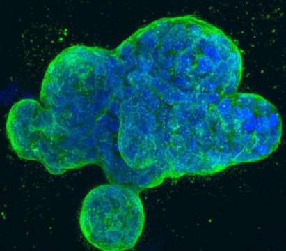
190103133648_1_540x360
This is a three-dimensional culture of human breast cancer cells, with DNA stained blue and a protein in the cell surface membrane stained green. Credit: National Institutes of Health

Adenosine deaminases are a family of three enzymes encoded by the ADAR genes, which stand for adenosine deaminase acting on RNA. They regulate gene expression by modifying nucleotides within double stranded RNA molecules, serving as fundamental editors in the development of new stem cells.

The enzyme, however, is also activated in cancers as diverse as liver, breast and leukemia. A research team led by senior author Catriona Jamieson, MD, PhD, deputy director of the Sanford Stem Cell Clinical Center and deputy director of the UC San Diego Moores Cancer Center, found that the normal functions of the ADAR1 enzyme are hijacked by pre-malignant cells, leading to a cascade of molecular consequences that promote malignant transformation, dormant cancer stem cell generation and resistance to treatment.
"We were able to illuminate the abilities of ADAR1 to 'hyper-mutate' tumor suppressor RNAs in leukemia and, at the same time, edit the microRNA aimed at targeting the tumor suppressor RNA. This enzyme turns on cancer resistance via a domino effect on RNA instead of DNA," said Qingfei Jiang, PhD, Assistant Project Scientist, Jamieson's lab.
Jamieson characterized RNA editing as tweaking basic genetic blueprints, not fundamentally rewriting them. Nonetheless, the results might be dramatic. "One result of detection of malignant RNA editing could be exposing dormant cancer stem cells that often escape therapies that target dividing cells, which leads to therapeutic resistance and disease relapse, and also highlight ADAR as a potentially tractable target for cancer stem cell elimination," said Jamieson.
Researchers from different part of the world are invited to submit abstract on their unpublished latest research at our upcoming conference Cell Tissue Science 2019 which is focused on the complications and consequences of Stem CellRegenerative MedicineStem Cell TherapyCancer Cell Biology,Technical Advancements in cancer treatment and many more. We as committee members of the conference welcome you to be a part of the conference “ 12th World Congress on Cell & Tissue Science” in Singapore on March 11-12, 2019
You can submit your abstract on Session or Track : 09. Stem Cells Biology With Regards to Christmas and New Year Celebration we are providing a special discount of 30% on all Registration Categories for more information please  visit by Click Here