Showing posts with label Cancer. Show all posts

Saturday, 1 June 2019

How prostate cancer cells mimic bone when they metastasize


Prostate cancer often becomes lethal as it spreads to the bones, and the process behind this deadly feature could potentially be turned against it as a target for bone-targeting radiation and potential new therapies.

Study published online in the journal PLOS ONE, Duke Cancer Institute researchers describe how prostate cancer cells develop the ability to mimic bone-forming cells called osteoblasts, enabling them to proliferate in the bone microenvironment.

Attacking these cells with radium-233, a radioactive isotope that selectively targets cells in these bone metastases, has been shown to prolong patients' lives. But a better understanding of how radium works in the bone was needed.

The mapping of this mimicking process could lead to a more effective use of radium-233 and to the development of new therapies to treat or prevent the spread of prostate cancer to bone.
"Given that most men who die of prostate cancer have bone metastases, this work is critical to helping understand this process," said lead author Andrew Armstrong, Director of Research at the Duke Cancer Institute Center for Prostate and Urologic Cancers.
The research team enrolled a small study group of 20 men with symptomatic bone-metastatic prostate cancer. When analyzing the circulating tumor cells from study participants, they found that bone-forming enzymes appeared to be expressed commonly, and that genetic alterations in bone forming pathways were also common in these prostate cancer cells.

They validated these new genetic findings in a separate multicenter trial involving a larger group of more than 40 men with prostate cancer and bone metastases.

Following treatment with radium-223, the researchers found that the radioactive isotope was concentrated in bone metastases, but tumor cells still circulated and cancer progressed within six months of therapy. The researchers found a range of complex genetic alterations in these tumor cells that likely enabled them to persist and develop resistance to the radiation over time.
"Osteomimicry may contribute in part to how prostate cancer spreads to bone, but also to the uptake of radium-223 within bone metastases and may thereby enhance the therapeutic benefit of this bone targeting radiotherapy," Armstrong said.
He said by mapping this lethal pathway of prostate cancer bone metastasis, the study points to new targets and thus critical areas of research into designing better tumor-targeting therapies.
An important announcement regarding our upcoming conference 12th World Congress on Cell & Tissue Science (Cell Tissue Science 2019) scheduled on September 13-14,2019 in Singapore. You can also present your latest research at the different topics such as Cancer Cell Biology, Stem Cell & its applications and many more along with other distinguished professors, doctors and researchers from all over the world.
If interested kindly proceed with submitting your abstract and latest biography along with a photography to our online abstract submission page given below: Link for submission: Click Here

Wednesday, 23 January 2019

New nanoparticle targets tumor-infiltrating immune cells, flips switch


Immunotherapy's promise in the fight against cancer drew international attention after two scientists won a Nobel Prize this year for unleashing the ability of the immune system to eliminate tumor cells.

But their approach, which keeps cancer cells from shutting off the immune system's powerful T-cells before they can fight tumors, is just one way to use the body's natural defenses against deadly disease. A team of Vanderbilt University bioengineers today announced a major breakthrough in another: penetrating tumor-infiltrating immune cells and flipping on a switch that tells them to start fighting. The team designed a nanoscale particle to do that and found early success using it on human melanoma tissue.
"Tumors are pretty conniving and have evolved many ways to evade detection from our immune system," said John T. Wilson, Assistant Professor of Chemical and Biomolecular Engineering and Biomedical Engineering. "Our goal is to rearm the immune system with the tools it needs to destroy cancer cells.Checkpoint blockade has been a major breakthrough, but despite the huge impact it continues to have, we also know that there are a lot of patients who don't respond to these therapies. We've developed a nanoparticle to find tumors and deliver a specific type of molecule that's produced naturally by our bodies to fight off cancer."
That molecule is called cGAMP, and it's the primary way to switch on what's known as the stimulator of interferon genes (STING) pathway: a natural mechanism the body uses to mount an immune response that can fight viruses or bacteria or clear out malignant cells. Wilson said his team's nanoparticle delivers cGAMP in a way that jump-starts the immune response inside the tumor, resulting in the generation of T-cells that can destroy the tumor from the inside and also improve responses to checkpoint blockade.

While the Vanderbilt team's research focused on melanoma, their work also indicates that this could impact treatment of many cancers, Wilson said, including breast, kidney, head and neck, neuroblastoma, colorectal and lung cancer.

His findings appear today in a paper titled "Endosomolytic Polymersomes Increase the Activity of Cyclic Dinucleotide STING Agonists to Enhance Cancer Immunotherapy" in the journal Nature Nanotechnology.

Daniel Shae, a Ph.D. student on Wilson's team and first author of the manuscript, said the process began with developing the right nanoparticle, built using "smart" polymers that respond to changes in pH that he engineered to enhance the potency of cGAMP. After 20 or so iterations, the team found one that could deliver cGAMP and activate STING efficiently in mouse immune cells, then mouse tumors and eventually human tissue samples.
"That's really exciting because it demonstrates that, one day, this technology may have success in patients," Shae said.
Researchers from different part of the world are invited to submit abstract on their unpublished latest research at our upcoming conference Cell Tissue Science 2019 which is focused on the complications and consequences of Stem CellRegenerative MedicineStem Cell Therapy, Cancer Cell Biology,Technical Advancements in cancer treatment and many more. We as committee members of the conference welcome you to be a part of the conference “ 12th World Congress on Cell & Tissue Science” in Singapore on March 11-12, 2019
You can submit your abstract on Session or Track : 08. Advancement in Cancer Treatments

Thursday, 13 December 2018

Unlocking the secrets of how cells communicate offers insights into treating diseases


2D images, left, and the 3D structure, right, of the gap junction communication channel from the eye lens, resolved to near-atomic resolution by the Reichow Lab using cryo-electron microscopy.
Credit: Reichow Lab | Portland State University
Portland State University researchers have made a significant breakthrough by developing the 3-D structure of proteins from inside the eye lens that control how cells communicate with each other, which could open the door to treating diseases such as cataracts, stroke and cancer.

The PSU research team, led by chemistry professor Steve Reichow, used a multimillion-dollar microscope and a novel technique developed by three Nobel Prize-winning biophysicists called cryo-electron microscopy (Cryo-EM) to view membrane protein channels or transportation tunnels in cell walls -- at the atomic level. This allowed Reichow's team, whose research is supported by the National Institutes of Health, to create a 3-D image of the membrane channel to better understand the processes involved in cell-to-cell communication.

Portland State researchers used Cryo-EM a microscope technique that freezes biomolecules in mid-movement and takes ultra-high-resolution images and computer modeling to see the 3-D structure of gap junction proteins that had been isolated from eye lenses. Gap junctions are tiny channels that allow neighboring cells to communicate with one another and are found in many places throughout the body.

Their findings published in the scientific Journal Nature -- showed for the first time how gap junctions selectively pass or block chemical information. Until now, it was not known how these channels would allow certain messages to pass between cells while specifically blocking others.

Reichow said the detailed images may open the door to understanding, and potentially treating, different types of diseases that are associated with the loss of function in cellular communication involving gap junctions such as cataracts, cardiac arrhythmia, stroke and certain cancers.
"Currently there's no drug on the market today that can specifically block or activate gap junction proteins," Reichow said. "But our discovery may one day pave the way for development of pharmaceuticals that can control heart disease or other diseases that are associated with the malfunctions or mis-regulations of these protein channels."
Now that the Reichow Lab has established a method for characterizing the proteins, they are trying to understand the nuances of how the body uses gap junctions differently across tissues and organs.
We welcome researchers from different part of the to submit abstract on their latest research at our upcoming conference Cell Tissue Science 2019 which is mainly focuses on the complications the consequences of Stem CellRegenerative MedicineStem Cell TherapyCancer Cell Biology , Technical Advancements in cancer treatment and many more.We welcome you to the our upcoming conference “ 12th World Congress on Cell & Tissue Science” . 
You can submit your abstract on Session or Track : 09. Stem Cells and its Applications

Wednesday, 5 December 2018

Novel DNA nanoplatform delivers anticancer agents to multidrug-resistant tumors


A tailored DNA nanoplatform carries chemotherapeutic drugs and RNA interference toward multidrug-resistant tumors

One of the most successful techniques to combat multidrug resistance in cancer cells is the downregulation of those genes responsible for drug resistance. Chinese scientists have now developed a nanoplatform that selectively delivers small hairpin RNA transcription templates and chemotherapeutics into multidrug-resistant tumors. A deadly cocktail of gene-silencing elements and chemotherapeutic drugs effectively and selectively kills cells, they reported in the Journal Angewandte Chemie. The nanoplatform was assembled using established DNA origami techniques.

Multidrug-resistant cancer cells often remove potent drugs from the cell before they can become effective. As several genes for proteins that perform this job are known, scientists attempt to interfere on the gene expression level, which is possible with RNA interference (RNAi) techniques: small RNAi strands combine with messenger RNA and inhibit transcription. However, RNA transcription templates must be delivered and released into the cytoplasm of the cell, and at the same time, a potent drug must be present to kill the cell.

Baoquan Ding at the National Center for Nanoscience and Technology, Beijing, China, and his colleagues have now designed and built a platform that includes every item needed to intrude into tumor cells and release gene-silencing elements and chemotherapeutic drugs. They built the platform using the DNA origami technique, which allows the construction of nanosized DNA objects in multiple, and even very complicated shapes. In this case, the scientists constructed a relatively simple DNA origami structure, which self-assembled into a triangular nanoplatform with various sites to bind multiple functional units.

One of the key features of the platform was that it could include the hydrophobic potent drug doxorubicin (DOX), a cytostatic that is especially useful against malign tumors. Here, DOX did not bind to the nanoplatform by any covalent linkage, but was loaded onto it through intercalation (which is the way DOX works in the cell: it intercalates into DNA, inhibiting transcription). Instead, what was covalently linked to the platform was the multiple gene silencing and cell-targeting site, which consisted of two linear small hairpin RNA transcription templates for RNAi and gene therapy, a cell-specific unit for specific recognition and insertion by the tumor cell, and a disulfide linkage to be cleaved by cellular glutathione.
The authors examined their multipurpose nanoplatform with an in vitro assay (on cell cultures) and by administering it into mice containing multidrug-resistant tumors. They found both a high and selective delivery and release rate of DOX and RNA transcription templates, and a high and selective tumor-killing efficiency. In addition, the multifunctional platform itself was not harmful to mice; however, filled with drugs and delivery sites, it was effective and deadly to multidrug-resistant tumors, the authors reported.

This research demonstrates what is possible in cancer therapy. The scientists have designed a nanostructure that not only specifically targets cancer cells, thus reducing severe side effects in chemotherapy, but also carries a drug and everything needed to suppress resistance in the cell when releasing the drug. And the platform itself is modifiable; adaption to other delivery strategies and other therapeutic components is easily possible, according to the authors.
We welcome researchers from different part of the to submit abstract on their latest research at our upcoming conference Cell Tissue Science 2019 which is mainly focuses on the complications the consequences of Stem CellRegenerative MedicineStem Cell TherapyCancer Cell Biology , Technical Advancements in cancer treatment and many more.We welcome you to the our upcoming conference “ 12th World Congress on Cell & Tissue Science” . 
You can submit your abstract on Session or Track : 8.Advancement in Cancer Treatment

Wednesday, 28 November 2018

Bone marrow-derived fibroblasts shown to promote breast cancer growth

A new study has revealed that breast cancer cells recruit bone marrow-derived fibroblasts to enhance their growth. It is hoped that the discovery will lead to the development of new treatments that target the cells in and around the tumor.

Many breast cancers show the presence of abundant fibroblasts from neighboring breast tissue and other body tissues.

Fibroblasts are typically associated with malignant solid tumors and are among the cell types that are not actually cancerous but enhance tumor progression.

For instance, fibroblasts within breast tumors secrete growth factors that increase the rate of proliferation of the cancer cells.

They also induce inflammation and tumor neovascularization (the formation of new blood vessels growing deep into the tumor).

Neovascularization of the tumor is essential to provide an adequate supply of oxygen and nutrients for its growth.

In the current experiment breast cancers in mice were shown to contain a high percentage of fibroblasts recruited from mesenchymal stromal cells (MSCs).

These cells originate in the bone marrow and show significant differences from other fibroblasts present in cancerous growths.

One important difference is the absence of an important cytokine, a protein called PDGFRα. The lack of PDGFRα, therefore, acts as a marker for such cells.

Bone marrow-derived fibroblasts are strong promoters of tumor neovascularization (the formation of new blood vessels to supply a tumor) because they secrete high amounts of the protein clusterin.

The presence of such fibroblasts was associated with faster tumor growth rates due to improved vascularization, compared to other breast tumors which contain only fibroblasts derived from surrounding breast tissue.

The same team discovered that human breast cancers also showed the presence of fibroblasts which were negative for PDGFRα.

When the tumor concentration of PDGFRα was lower, the tumors were significantly more likely to be fatal.

In effect, therefore, breast tumors that recruit bone marrow-derived fibroblasts have taken a very important step towards more rapid growth and distant spread.

Gaining insight into how these cells work could well lead to the emergence of new treatments which selectively target not just the cancer cells but also the bone marrow-derived fibroblasts that support tumor progression.
Our study shows that the recruitment of bone marrow-derived fibroblasts is important for promoting tumor growth, likely by enhancing blood vessel formation. Understanding the function of these cancer-associated fibroblasts could form the basis of developing novel therapeutic manipulations that cotarget bone marrow-derived fibroblasts as well as the cancer cells themselves."
Neta Eretz, Lead Author
We welcome researchers from different part of the to submit abstract on their latest research at our upcoming conference Cell Tissue Science 2019 which is mainly focuses on the complications the consequences of Stem CellRegenerative MedicineStem Cell TherapyCancer Cell Biology , Technical Advancements in cancer treatment and many more.We welcome you to the our upcoming conference “ 12th World Congress on Cell & Tissue Science” . 
You can submit your abstract on Session or Track : 7.Cancer Cell Biology

Tuesday, 27 November 2018

New lung cancer guides aim to drive better outcomes and care

wo new, national guides that aim to drive better outcomes and care for all people affected by lung cancer were released by Cancer Australia. Lung cancer is the leading cause of death from cancer in Australia with an estimated 25 people dying every day from the disease in 2018.
All lung cancer patients, regardless of where they live, should have the benefit of the best care and have the best possible experience. These resources distill the essential elements of what that care should be and how to deliver it,” Dr Helen Zorbas, CEO Cancer Australia
Separate resources have been designed for health professionals and consumers, which share the goal of achieving optimal patient outcomes.
Delivering best practice lung cancer care for health professionals contains evidence-based, best practice information, strategies, tools and resources to support clinicians in providing consistent, high quality care for people affected by lung cancer.
I encourage all those involved in lung cancer care in Australia to implement and use these guides as part of a national coordinated effort to improve lung cancer outcomes.” Dr Zorbas
The resources build on the Lung Cancer Framework: Principles for Best Practice Lung Cancer Care in Australia, which support the uptake and use of the Principles for best practice care: patient-centred care; timely access to evidence-based pathways of care; multi-disciplinary care; coordination, communication and continuity of care; and data-driven improvements in lung cancer care.

To empower patients to actively participate in their own care, Getting the best advice and care, a guide for those affected by lung cancer provides information about what the principles will mean to them and what they can do better engage with health care professionals and make informed evidence-based decisions.
Dr Zorbas said the guides articulate the clear benefits to patients of implementing each of the principles –from improving survival to reducing anxiety and depression.

Releasing these resources during Lung Cancer Awareness Month serves as a timely reminder that anyone experiencing symptoms such as a persistent cough lasting 3 weeks or more, coughing up blood, a chest infection that won’t go away, or a changed cough should visit a doctor.

Although lung cancer mostly occurs in people over 60 years, it can affect people of any age, regardless of whether they smoke. It is estimated that in 2018, 12,741 people will be diagnosed with lung cancer in Australia.

The development of Lung Cancer Framework: Principles for Best Practice Lung Cancer Care in Australia and these new resources by Cancer Australia was guided by systematic and exacting reviews of the highest quality evidence and incorporates learnings and experiences from people affected by lung cancer. A national demonstration project was undertaken, along with consultation with health professionals and consumers, supported by Cancer Australia’s Lung Cancer Advisory Group.
We welcome researchers from different part of the to submit abstract on their latest research at our upcoming conference Cell Tissue Science 2019 which is mainly focuses on the complications the consequences of Stem CellRegenerative MedicineStem Cell TherapyCancer Cell Biology , Technical Advancements in cancer treatment and many more.We welcome you to the our upcoming conference “ 12th World Congress on Cell & Tissue Science” . 
You can submit your abstract on Session or Track : 8.Advancement in Cancer Treatment

Friday, 23 November 2018

Nutritional supplement can slow cancer growth and enhance effects of chemotherapy


Mannose sugar, a nutritional supplement, can both slow tumor growth and enhance the effects of chemotherapy in mice with multiple types of cancer.
This lab study is a step towards understanding how mannose could be used to help treat cancer.

The results of the study, which was funded by Cancer Research UK and Worldwide Cancer Research, are published in Nature.

Tumors use more glucose than normal, healthy tissues. However, it is very hard to control the amount of glucose in your body through diet alone. In this study, the researchers found that mannose can interfere with glucose to reduce how much sugar cancer cells can use.
Professor Kevin Ryan, lead author from the Cancer Research UK Beatson Institute, said: "Tumors need a lot of glucose to grow, so limiting the amount they can use should slow cancer progression. The problem is that normal tissues need glucose as well, so we can’t completely remove it from the body. In our study, we found a dosage of mannose that could block enough glucose to slow tumor growth in mice, but not so much that normal tissues were affected. This is early research, but it is hoped that finding this perfect balance means that, in the future, mannose could be given to cancer patients to enhance chemotherapy without damaging their overall health.”
The researchers first examined how mice with pancreatic, lung or skin cancer responded when mannose was added to their drinking water and given as an oral treatment. They found that adding the supplement significantly slowed the growth of tumors and did not cause any obvious side effects.

To test how mannose could also affect cancer treatment, mice were treated with cisplatin and doxorubicin - two of the most widely used chemotherapy drugs. They found that mannose enhanced the effects of chemotherapy, slowing tumor growth, reducing the size of tumors and even increasing the lifespan of some mice.

Professor Kevin Ryan added:"Our next step is investigating why treatment only works in some cells, so that we can work out which patients might benefit the most from this approach. We hope to start clinical trials with mannose in people as soon as possible to determine its true potential as a new cancer therapy.”
Mannose is sometimes used for short periods to treat urinary tract infections, but its long-term effects have not been investigated. It’s important that more research is conducted before mannose can be used in cancer patients.
Martin Ledwick, Cancer Research UK’s head nurse, said:"Although these results are very promising for the future of some cancer treatments, this is very early research and has not yet been tested in humans. Patients should not self-prescribe mannose as there is a real risk of negative side effects that haven’t been tested for yet. It’s important to consult with a doctor before drastically changing your diet or taking new supplements.”
We welcome researchers from different part of the to submit abstract on their latest research at our upcoming conference Cell Tissue Science 2019 which is mainly focuses on the complications the consequences of Stem CellRegenerative MedicineStem Cell TherapyCancer Cell Biology , Technical Advancements in cancer treatment and many more.We welcome you to the our upcoming conference “ 12th World Congress on Cell & Tissue Science” . 
You can submit your abstract on Session or Track : 8.Advancement in Cancer Treatment

Friday, 16 November 2018

People with rare cancers can benefit from genomic profiling


New research has shown that many Australians with rare cancers can benefit from genomic profiling. The findings of the patient-driven trial are being presented today at the Clinical Oncology Society of Australia Annual Scientific Meeting and could result in dramatic changes to the way those with rare cancers are diagnosed and treated. 

The initial data from the pilot study for Nominator Trial is being presented by Professor Clare Scott from the Walter and Eliza Hall Institute of Medical Research and Peter MacCallum Cancer Centre, and was funded in part by Rare Cancers Australia

The data shows that genomic profiling provides meaningful information that influences diagnosis and treatment in approximately 50 percent of people with rare cancers. 20 percent of those tested got a new treatment plan as a result and 6 percent of participants were given a new diagnosis. 

The aim of the national initiative is to trial the use of genomic testing to match rare cancers to cancer treatments. Testing is used to identify molecular features of the cancer or genetic mutations that can be targeted with existing treatments used in other cancer types with the same characteristics.

While genomic testing is becoming increasingly used in other cancer types, this is one of the first Australian studies of its kind to look at the potential benefits for those with rare cancers, which have very low survival rates.
Professor Clare Scott says the initial pilot data is exciting and proves that there is a current unmet need."The treatment options for Australians with rare cancers are currently extremely limited and this ultimately leads to poor survival rates. Research has also typically been restricted because of the challenges of finding enough of each type of cancer patient to design appropriate clinical trials."Australians in this trial came to us after they had exhausted all their options. The cancers they had are extremely rare - the chances of being diagnosed with these cancer types are often around one in a million."Using genomic profiling we were able to uncover new information that gave many patients new treatment options - and ultimately, new hope."
Professor Scott says."In one case we were able to identify that a rare heart tumour actually had a genetic profile most closely resembling a melanoma. Using that information we were able to get access to the latest treatments that are benefiting melanoma patients - which we hope will provide better outcomes for this patient."
The Nominator Pilot Study results released today included 36 patients. The two-year study will eventually include 100 patients and will lay the groundwork for other national initiatives looking into genomic profiling across a range of cancer types. Professor Phyllis Butow, President, Clinical Oncology Society of Australia said one of the impressive things about the study was that it was driven by Australians directly affected by rare cancers.
"Around 52,000 Australians are diagnosed with rare or less common cancers each year. Those directly affected by the disease, led by Kate and Richard Vines from Rare Cancers Australia, helped call for and fund this research, so it's great to see these initial promising results being presented to cancer experts from across the country." 
We welcome researchers from different part of the to submit abstract on their latest research at our upcoming conference Cell Tissue Science 2019 which is mainly focuses on the complications the consequences of Stem CellRegenerative MedicineStem Cell TherapyCancer Cell Biology , Technical Advancements in cancer treatmentand many more.We welcome you to the our upcoming conference “ 12th World Congress on Cell & Tissue Science” .  You can submit your abstract on Session or Track :08- Advancement in Cancer Treatments

Wednesday, 14 November 2018

Scientists uncover hidden estrogen receptors in epithelial breast cells


Estrogens are hormones that play central roles in the development and the physiology of the breast, but also are involved in breast cancer. Like all hormones, estrogens exert their biological effects by binding to dedicated receptors in the target cell.

Scientists led by Cathrin Brisken at EPFL have now uncovered that half of the luminal epithelial breast cells that appear not to express the estrogen receptor actually express it at low levels. Publishing in Nature Communications, they show that different parts of the estrogen receptor play different roles in the luminal breast cells that give rise to cancer. Depending on whether a cell has low or high levels of the estrogen receptor, the hormone-dependent or the hormone-independent activities are more or less important for its function.

In addition, the researchers found that the action of the estrogen receptor is biphasic: it stimulates the expansion and growth of breast cells in young mice but inhibits it during pregnancy.
The discovery has immediate implications for the role of ERα in the development of breast cancer. "This begs the question whether these ER-pseudo-negative breast cells will ultimately turn into estrogen receptor-positive or -negative breast cancers," says Cathrin Brisken.
We welcome researchers from different part of the to submit abstract on their latest research at our upcoming conference Cell Tissue Science 2019 which is mainly focuses on the complications the consequences of Stem CellRegenerative MedicineStem Cell TherapyCancer Cell Biology , Technical Advancements in cancer treatmentand many more.We welcome you to the our upcoming conference “ 12th World Congress on Cell & Tissue Science” . 
You can submit your abstract on Session or Track :1.Cell Biology

Tuesday, 13 November 2018

New methods to identify AD drug candidates with anti-aging properties


Long thought to suppress cancer by slowing cellular metabolism, the protein complex AMPK also seemed to help some tumors grow, confounding researchers. Now, Salk Institute researchers have solved the long-standing mystery around why AMPK can both hinder and help cancer.

The lab of Salk Professor Reuben Shaw showed that late-stage cancers can trigger AMPK's cellular recycling signal to cannibalize pieces of the cell, supplying large lung tumors with the nutrients they need to grow. The work was published in  Cell Metabolism suggests that blocking AMPK in some conditions could stop the growth of advanced tumors in the most common type of lung cancer.
"Our study shows that the same dysfunction in a genetic circuit that causes non-small-cell lung cancer to begin with is necessary for more mature tumor cells to survive when they don't have enough nutrients," says Shaw, Director of the Salk Cancer Center. "It's exciting because not only does it solve a genetic 'whodunnit,' but it also points to a potential new therapeutic target for a cancer that is often diagnosed very late."
AMPK acts as a fuel gauge for the cell, overseeing energy input and output to keep the cell running smoothly. Similar to a car sensor flashing a low-gas signal or turning off a vehicle's AC to save energy, AMPK slows down cell growth and changes the cell's metabolism if the cell's fuel (nutrients) is low. Previously, Shaw discovered that AMPK could halt tumors' revved-up metabolism, as well as restore normal function to the liver and other tissues in diabetics.
"We found that tumors grew much more slowly when AMPK was not present," says Research Associate Lillian Eichner."That means that AMPK is not always functioning as a tumor suppressor, as we originally thought."
The team analyzed which genes in tumor cells from the same mouse models were being activated under various conditions. One gene that was particularly active was Tfe3, which is known to activate cellular recycling. It turned out that when tumors became large enough that cells in the middle were too far from easy access to nutrients, AMPK signaled Tfe3 to initiate recycling of cellular materials as nutrients--effectively cannibalizing pieces of the cell--for the tumor to use.
"Previously we were focused on how we could activate AMPK," says Eichner. "Now that we've identified this mechanism, we can shift to how to inhibit it in certain cancers."
Shaw, who holds the William R. Brody Chair, adds, "We're excited because more advanced tumors seem to rely on AMPK to survive, and understanding this mechanism means we may be able to treat them."
We welcome researchers from different part of the to submit abstract on their latest research at our upcoming conference Cell Tissue Science 2019 which is mainly focuses on the complications the consequences of Stem CellRegenerative MedicineStem Cell TherapyCancer Cell Biology , Technical Advancements in cancer treatmentand many more.We welcome you to the our upcoming conference “ 12th World Congress on Cell & Tissue Science” . 
You can submit your abstract on Session or Track : 16.Anti-Aging Medicine

Monday, 12 November 2018

Realizing the potential of gene therapy for neurological disorders


Promising findings from preclinical animal studies show the potential of gene therapy for treating incurable neurological disorders. In new research presented today, scientists successfully used gene therapy to slow the progression and improve symptoms of disorders such as amyotrophic lateral sclerosis and Parkinson's disease.

Gene therapy typically employs an inactivated virus to carry new genetic cargo into cells, altering specific genes to treat or prevent a disease. Researchers might replace a mutated gene with a healthy copy of the gene, turn off a disease-causing gene, or add a new gene to the body to help fight a disease.

Although gene therapy is a promising treatment option for a limited number of conditions, including certain cancers, the technique is still experimental for most diseases, with ongoing research to ensure they will be safe and effective in human patients. Animal studies are a key part of the process by which an experimental gene therapy treatment goes to clinical trial.

Research new findings show that:
  • A new technique allows gene therapy to be delivered to the entire spinal cord in mice (M. Bravo Hernandez, abstract 208.10).
  • Gene therapy safely and effectively extends life and improves motor function in a mouse model of ALS (Gretchen Thomsen, abstract 208.16).
  • Gene therapy slows the progression of neuronal loss in a mouse model of inherited Parkinson's disease (Jose L. Lanciego, abstract 292.01).
  • Gene therapy shows promise in a mouse model of Batten disease, a childhood neurodegenerative disease (Shibi Likhite, abstract 355.01).
"Gene therapy holds the promise to transform the lives of patients with incurable neurological diseases," said Fredric Manfredsson, PhD, of Michigan State University. "The research presented today represents important and exciting steps toward being able to prevent and treat disorders that currently have no cure, such as Parkinson's disease and Alzheimer's disease."
We welcome researchers from different part of the to submit abstract on their latest research at our upcoming conference Cell Tissue Science 2019 which is mainly focuses on the complications the consequences of Stem CellRegenerative MedicineStem Cell TherapyCancer Cell Biology , Technical Advancements in cancer treatmentand many more.We welcome you to the our upcoming conference “ 12th World Congress on Cell & Tissue Science” . 
You can submit your abstract on Session or Track :2.Cellular and Gene Therapy

Friday, 12 October 2018

Redrawing the structure of an immune system protein

Researchers have revealed the structure of an essential immune protein, creating future possibilities to develop more effective medicines for a range of illnesses from cancer to neurological diseases. University of Tokyo researchers made this discovery with computerized image analysis and modern electron microscope imaging.
Cell Tissue Science2019
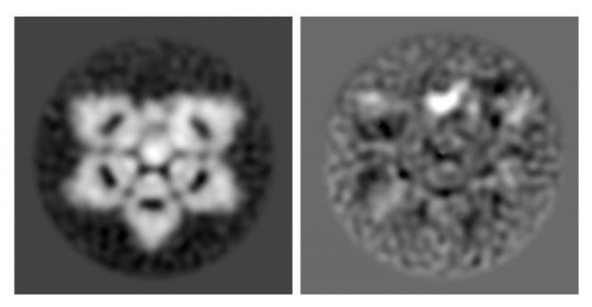
Electron microscopes and modern computer image processing allowed University of Tokyo researchers to uncover the correct structure of a protein essential in the immune system, IgM, and a much smaller protein that is bound inside, AIM. IgM is the larger, incomplete hexagon shape and AIM is the smaller broad bean shape inside the wedge-shape gap. In the right-side image, AIM is more easily recognized as the bright white, broad-bean-shaped spot.
Credit: Image by Hiramoto et al., originally published in Science Advances, CC-BY
Researchers verified the structure of natural immunoglobulin M (IgM) protein, an important part of the immune system, using mouse and human versions of the protein. IgM is now understood to be shaped like an incomplete hexagon, or a pentagon with a wedge-shaped gap.

IgM is the first immune system protein that develops in the human fetus and remains the first responder to pathogens throughout life. The structure of IgM was first identified in 1969 as a "five-pointed, star-shaped table" and updated in 2009 to be a five-sided dome or "mushroom cap."
"The original IgM model was made by looking at a few individual molecules by hand with what today we think is a low-resolution microscope. Now we have clearer pictures and the computer can examine thousands of individual IgM molecules," said Miyazaki.
The 2018 discovery of the incomplete hexagon was actually a secondary interest for Miyazaki, originally a medical doctor who has built his research career studying a different protein called apoptosis inhibitor of macrophage (AIM).

Since identifying the correct shape of IgM, researchers now understand that inactive AIM is nestled inside the gap of the IgM incomplete hexagon. The structural connection between IgM and AIM means that drugs with the ability to regulate the release of AIM could be used to create AIM-based disease therapies.
"We can think of AIM as a fighter jet and IgM as the aircraft carrier ship. When other molecules activate the immune system, IgM releases AIM. The much smaller AIM protein goes around the body to clear away damaged cells and prevent disease," said Miyazaki.
Miyazaki identified AIM in 1999 while working at the Basel Institute for Immunology Institute in Switzerland. Its small size means AIM is easily eliminated from the body by the kidneys and excreted in urine, so staying bound within the larger IgM protects AIM from being removed before it is needed.

AIM is a common molecule in the bloodstream, but it is only active when the body develops a disease. AIM is known to be important for preventing obesity, fatty liver disease, hepatocellular carcinoma (liver cancer), multiple sclerosis (MS), fungus-induced peritonitis (inflammation of the abdominal wall membrane), and acute kidney injury.

The incomplete hexagon structure is still only a 2D understanding of IgM structure. Miyazaki and his team continue to do additional analysis and hope to report the 3D structure of IgM soon.
We welcome researchers from different part of the to submit abstract on their latest research at our upcoming conference Cell Tissue Science 2019 which is mainly focuses on the complications the consequences of Stem CellRegenerative MedicineStem Cell TherapyCancer Cell Biology , Technical Advancements in cancer treatment and many more.We welcome you to the our upcoming conference “ 12th World Congress on Cell & Tissue Science” . For more info visit :Cell Tissue Science 2019

Thursday, 4 October 2018

Diet affects the breast microbiome in mammals

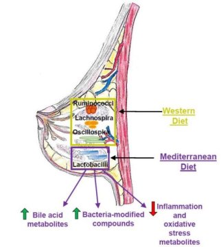
Diet influences the composition of microbial populations in the mammary glands of nonhuman primates. Specifically, a Mediterranean diet increased the abundance of probiotic bacteria previously shown to inhibit tumor growth in animals.
Cell Tissue Science 2019
This image shows how diet plays a critical role in determining microbiota populations in tissues outside the gut, such as the mammary gland. Credit: Katherine Cook

"We showed for the first time that breast-specific microbiome populations are significantly affected by diet, and this was in a well-established nonhuman primate model of women's health, increasing the likelihood that these findings will be important for human health," says Carol Shively, Wake Forest School of Medicine. "The breast microbiome is now a target for intervention to protect women from breast cancer."
Diet has been extensively studied as a lifestyle factor that could influence breast cancer development. Breast cancer risk in women is increased by consumption of a high-fat Western diet full of sweets and processed foods but reduced by a healthy Mediterranean diet consisting of vegetables, fish, and olive oil. Intriguingly, a recent study in humans revealed that malignant breast tumors have a lower abundance of Lactobacillus bacteria compared to benign lesions, suggesting that microbial imbalances could contribute to breast cancer.
"However, it was unknown what possible factors could modulate the breast tissue microbiome," says Katherine Cook, Wake Forest School of Medicine. "Diet is a strong influencer on the gut microbiome, so we decided to test the hypothesis that diet can impact mammary gland microbiota populations."
To address this question, Shively and Cook used macaque monkeys because the animals mimic human breast biology and have been used to study breast cancer risk. One advantage over human studies is that the food intake of the monkeys can be carefully controlled for a prolonged period of time, increasing the chance of observing profound effects of diet.
The researchers assigned 40 adult female monkeys to receive either a Western or a Mediterranean diet for 31 months. The breast tissue of monkeys that consumed a Mediterranean diet had a 10-fold higher abundance of Lactobacillus, which is commonly used in probiotics and has been shown to decrease tumor growth in animals with breast cancer. The Mediterranean diet also increased levels of bile acid metabolites and bacterial-processed bioactive compounds that may decrease breast cancer risk.
Taken together, these results suggest that diet directly influences microbiome populations outside of the intestinal tract and could impact mammary gland health. But for now, it is not clear what impact these microbes or microbial-modified metabolites have on breast cancer risk.
Moving forward, the Cook lab plans to investigate the physiological impact of increased Lactobacillus in mammary gland tissue. They will also assess whether oral interventions such as fish oil or probiotic supplements can impact microbial populations in mammary glands and breast tumors. In addition, they are exploring the role of bacterial-modified bioactive compounds and bile acids on inflammation, breast cancer tumor growth, and therapeutic responsiveness.
"Our future studies are designed to validate the use of probiotics, fish oil, or antibiotics during neoadjuvant therapy to improve therapeutic outcomes," Cook says. "We hope to translate our studies into the clinic in the near future."
We encourage researchers all around the globe to submit abstract on their latest research at our upcoming conference Cell Tissue Science 2019 which is mainly focuses on the complications the consequences of Stem CellRegenerative MedicineStem Cell TherapyCancer Cell Biology , Technical Advancements in cancer treatment   and many more.We welcome you to the our upcoming conference “ 12th World Congress on Cell & Tissue Science” . For more info visit :Cell Tissue Science 2019

Saturday, 29 September 2018

Enzymes 'partner up' to accelerate cancer, aging diseases

A new study from molecular biologists at Indiana University has identified cellular processes that appear to supercharge both the growth and shrinkage of the chemical "caps" on chromosomes associated with aging, called telomeres.

The work, focused on two enzymes in yeast, could lead to new insights on stopping runaway cellular growth in cancer tumors, as well as the treatment of premature aging disorders such as progeria (aka "Benjamin Button disease").
"This work confirms that two specific enzymes are called helicases which are involved in telomere maintenance, and demonstrates they're even stronger in combination.This is significant since dysfunction in telomere maintenance has been found in 100 percent of cancers. Literally, 100 percent. So, it's very likely they play a role in the disease."said Matthew Bochman, Associate Professor, IU Bloomington College of Arts and Sciences 
Helicases are enzymes that unwind double-stranded DNA into a single stand for the purposes of replication, recombination and repair. In humans, the RecQ4 helicase functions similarly to the Hrq1 helicase in yeast. The Pif1 helicase is the same in both species.

In healthy people, telomeres shorten slowly over the lifespan as part of the natural aging process. In cancer cells, the brakes come off this process, so telomeres never grow shorter -- resulting in uncontrolled cellular replication. In people with premature aging disorders, telomeres rapidly shrink, resulting in death from "old age" in the late teenage years.

Mutations of the Pif1 helicase have been linked to several types of cancer, including common forms such as breast, ovarian and colon cancer. Mutations in the RecQ4 helicase have been linked to three different diseases associated with predispositions for cancer.

The study specifically found that Hrq1 and Pif1 are a "dynamic duo" that combine to create a "super inhibitor" or, under certain specific conditions, a "super stimulator" of telomere growth.
"Now, we can now really start to pick apart more about how these enzymes work together or alone in cancer cells," Bochman said.
The work may help scientists better understand whether certain cancers involve errors in DNA recombination, DNA repair or telomere maintenance or some other mechanistic problem. This, in turn, could lead to new ways to disrupt or harness these processes with drugs or other therapies.
 
We welcome researchers from different part of the to submit abstract on their latest research at our upcoming conference Cell Tissue Science 2019 which is mainly focuses on the complications the consequences of Stem CellRegenerative MedicineStem Cell TherapyCancer Cell Biology , Technical Advancements in cancer treatment and many more.We welcome you to the our upcoming conference “ 12th World Congress on Cell & Tissue Science” . For more info visit :Cell Tissue Science 2019

Friday, 28 September 2018

Understanding epilepsy in pediatric tumors

Pediatric brain tumors are characterized by frequent complications due to intractable epilepsy compared to adult brain tumors. However, the genetic cause of refractory epilepsy in pediatric brain tumors has not been elucidated yet, and it is difficult to treat patients because the tumors do not respond to existing antiepileptic drugs and debilitate children's development.

A Korean research team led by Professor Jeong Ho Lee, Graduate School of Medical Science and Engineering, Korea Advanced Institute of Science and Technology (KAIST) has recently identified a neuronal BRAF somatic mutation that causes intrinsic epileptogenicity in pediatric brain tumors.


180919133918_1_900x600
Preoperative and postoperative brain MRI (left panel), tumor H&E (right upper panel) and GFAP immunohistochemical (right lower panel) staining images from a patient with ganglioglioma (GG231) carrying the BRAFV600E mutation. The white arrow and the black arrowhead indicate the brain tumor and a dysplastic neuron, respectively.
Credit: KAIST

The research team studied patients' tissue diagnosed with ganglioglioma (GG), one of the main causes of tumor-associated intractable epilepsy, and found that the BRAF V600E somatic mutation is involved in the development of neural stem cells by using deep DNA sequencing. This mutation was carried out in an animal model to reproduce the pathology of GG and to observe seizures to establish an animal model for the treatment of epileptic seizures caused by pediatric brain tumors.

Using immunohistochemical and transcriptome analysis, they realized that the BRAF V600E mutation that arose in early progenitor cells during embryonic brain formation led to the acquisition of intrinsic epileptogenic properties in neuronal lineage cells, whereas tumorigenic properties were attributed to a high proliferation of glial lineage cells exhibiting the mutation. Notably, researchers found that seizures in mice were significantly alleviated by intraventricular infusion of the BRAF V600E inhibitor, Vemurafenib, a clinical anticancer drug.
The authors said, "Our study offers the first direct evidence that the BRAF somatic mutation arising from neural stem cells plays a key role in epileptogenesisin the brain tumor. This study also showed a new therapeutic target for tumor-associated epileptic disorders."
In collaboration with the KAIST startup company, SoVarGen, the research team is currently developing innovative therapeutics for epileptic seizures derived from pediatric brain tumors. This study was supported by the Suh Kyungbae Foundation (SUHF) and the Citizens United for Research in Epilepsy.
 
We encourage researchers all around the globe to submit abstract on their latest research at our upcoming conference Cell Tissue Science 2019 which is mainly focuses on the complications the consequences of Stem CellRegenerative MedicineStem Cell TherapyCancer Cell Biology , Technical Advancements in cancer treatment   and many more.We welcome you to the our upcoming conference “ 12th World Congress on Cell & Tissue Science” . For more info visit :Cell Tissue Science 2019

Wednesday, 19 September 2018

Heart attack: Substitute muscle thanks to stem cells

Scientists from University of Würzburg  have for the first time succeeded in generating beating cardiac muscle cells from special stem cells. They may provide a new approach for the treatment of heart attacks.
Myocardial infarction -- commonly known as a heart attack -- is still one of the main causes of death. According to the Federal Statistical Office, more than 49,00 people died of its consequences. And yet the mortality after heart attack has greatly decreased over the past decades: As compared to the early 1990s, it has more than halved until 2015, according to the German Society of Cardiology (DGK). The reasons for this include better prevention, therapy and rehabilitation.
A heart attack leaves scars
The problem is during each heart attack, some of the cardiac muscle tissue dies which is accompanied by more or less marked scarring. Attempts made over the past years to substitute the destroyed tissue by adequately functioning cardiac muscle using stem cells have not been as successful as expected.
Recent results of research scientists at the University of Würzburg (JMU) now show a novel approach for the treatment of myocardial infarction. The team around Professor Süleyman Ergün, , focuses on a special type of stem cells that it has discovered in vascular walls.
Help from intracardiac blood vessels
"We could demonstrate for the first time the presence of special stem cells in human vascular walls that have the ability to develop into beating cardiac muscle cells under culture conditions," explains Professor Ergün, Head of the Institute of Anatomy and Cell Biology, University of Würzburg. The scientists could also show that these "vascular wall-resident stem cells" exist also in the walls of intracardiac blood vessels, the so-called "coronary vessels," and are actually activated to respond when a heart attack happens.
The problem so far was that in the event of an infarction these stem cells had no chance to develop into cardiac muscle cells as desired: "Our studies have shown that these cells are integrated into the scar tissue and thus lose their ability to transform into cardiac muscle cells," explains the scientist. Nevertheless the results give cause for hope: "Our results provide a new approach, in that it may be possible to therapeutically manipulate the behavior of the stem cells in the intracardiac vascular walls so that they are stimulated into regenerating the destroyed cardiac muscle tissue" says Dr. Ergün.
A novel therapy approach
Therapists are convinced that if a timely and therapeutically effective control of the newly discovered stem cells from the intracardiac blood vessels were actually possible, it would mean a huge step forward in the treatment of cardiovascular diseases. At the same time, it offers a chance to significantly reduce the therapeutic cost of these diseases. However, the scientists' findings are still limited to studies on experimental animals and in the lab. Further studies are therefore required to deepen the findings obtained before they can be used on humans.
 
We welcome researchers from different part of the to submit abstract on their latest research at our upcoming conference Cell Tissue Science 2019 which is mainly focuses on the complications the consequences of Stem CellRegenerative MedicineStem Cell TherapyCancer Cell Biology , Technical Advancements in cancer treatment and many more.We welcome you to the our upcoming conference “ 12th World Congress on Cell & Tissue Science” . For more info visit :Cell Tissue Science 2019

New method promises fewer side effects from cancer drugs

Researchers from Faculty of Science - University of Copenhagen have suggested that Protein research is one of the hottest areas in medical research because proteins make it possible to develop far more effective pharmaceuticals for the treatment of diabetes, cancer and other illnesses.
Proteins have incredibly complex chemical structures that make them difficult to modify. As a result, researchers have been looking for a tool to modify them more precisely, without increasing a drug's side-effects.
"We often run the risk of not being approved by health authorities because protein-based drugs lack precision and may have side-effects. Among other things, this is because of the serious limitations with the tools that have been used up until now," according to Professor Knud J. Jensen, University of Copenhagen's Department of Chemistry.
Together with his research colleague, Sanne Schoffelen, he has developed a new protein-modifying method that promises fewer side-effects and could be pivotal in furthering the development of protein-based pharmaceuticals.
Protein structure is like an intricate ball of yarn
Researchers call the method "His-tag acylation." Among other things, it makes it possible to add a toxic molecule to proteins that can attack sick cells in a cancer-stricken body without attacking healthy ones.
"Proteins are like a ball of yarn, a long thread of amino acids, which are turned up. This method allows us to precisely target these intricate structures, as opposed to making uncertain modifications when we don't know what is being hit within the ball of yarn. In short, it will help produce drugs where we can be far more confident about where modifications are being made, so that side effects can be minimized in the future," says Knud J. Jensen.
Modified proteins must target precisely
The fact that His-tag acylation can accurately target these complex yarn-like protein structures also makes it possible to produce drugs with entirely new characteristics.
For example, researchers can now attach a fluorescent molecule to proteins in such a way that a microscope can be used to track a protein's path through cells. The primary function of these proteins is to transport cancer fighting molecules around to sick cells, so it is important to carefully follow their path throughout the body in order to safely produce medications that don't have unintended side-effects.
 
We welcome researchers all around the globe to submit abstract on their latest research at our upcoming conference Cell Tissue Science 2019 which is mainly focuses on the complications the consequences of Stem CellRegenerative MedicineStem Cell TherapyCancer Cell Biology , Technical Advancements in cancer treatment and many more.We welcome you to the our upcoming conference “ 12th World Congress on Cell & Tissue Science” . For more info visit :Cell Tissue Science 2019