Showing posts with label Research. Show all posts

Saturday, 15 June 2019

'Virtual biopsy' device to detect skin tumors


Using sound vibrations and pulses of near-infrared light, a Rutgers University scientist has developed a new "virtual biopsy" device that can quickly determine a skin lesion's depth and potential malignancy without using a scalpel.

The ability to analyze a skin tumor non-invasively could make biopsies much less risky and distressing to patients, according to a report in Wiley Online Library. Currently, physicians who perform surgical biopsies often don't know the extent of a lesion and whether it will be necessary to refer the patient to a specialist for extensive tissue removal or plastic surgery until surgery has already begun.

The first-of-its-kind experimental procedure, called vibrational optical coherence tomography (VOCT), creates a 3-D map of the legion's width and depth under the skin with a tiny laser diode. It also uses soundwaves to test the lesion's density and stiffness since cancer cells are stiffer than healthy cells. An inch-long speaker applies audible soundwaves against the skin to measure the skin's vibrations and determine whether the lesion is malignant.
"This procedure can be completed in 15 minutes with no discomfort to the patient, who feels no sensation from the light or the nearly inaudible sound. It's a significant improvement over surgical biopsies, which are invasive, expensive and time consuming," said Frederick Silver, a professor of pathology and laboratory at Rutgers Robert Wood Johnson Medical School.
The study found that a prototype VOCT device, which awaits FDA approval for large-scale testing, is able to accurately distinguish between healthy skin and different types of skin lesions and carcinomas. The researchers tested the device over six months on four skin excisions and on eight volunteers without skin lesions. Further studies are needed to fine-tune the device's ability to identify a lesion's borders and areas of greatest density and stiffness, which would allow physicians to remove tumors with minimally invasive surgery.

An important announcement regarding our upcoming conference 12th World Congress on Cell & Tissue Science (Cell Tissue Science 2019) scheduled on September 13-14,2019 in Singapore. You can also present your latest research at the different topics such as Cancer Cell Biology, Stem Cell & its applications and many more along with other distinguished professors, doctors and researchers from all over the world.
If interested kindly proceed with submitting your abstract and latest biography along with a photography to our online abstract submission page given below: Link for submission: Click Here
Source: https://www.sciencedaily.com/releases/2019/06/190613103129.htm

Friday, 7 June 2019

New genes out of nothing


One key question in evolutionary biology is how novel genes arise and develop. Swedish researchers now show how new genes and functions that are advantageous to bacteria can be selected from random DNA sequences. The results are presented in the scientific journal mBio.

How do new genes and functional proteins arise and develop? This is one of the most fundamental issues in evolutionary biology. Two different types of mechanism have been proposed: (1) new genes with novel functions arise from existing genes, and (2) new genes and proteins evolve from random DNA sequences with no similarity to existing genes and proteins.

In the present study, the researchers explored the latter type of mechanism: evolution of new genes and proteins from randomised DNA sequences as its called as de novo evolution. It is fairly easy to understand that when a gene already exists, it can be modified and acquire a new function. But how does "nothing" turn into a function affording a small advantage that is favoured by natural selection?

The raw material for the experiment was an big library of some 500 million randomised gene sequences, from which peptide sequences with a biological function were identified. In the experiment, random gene sequences were placed on a plasmid and overexpressed. The scientists then investigated whether they could give bacteria a specific, defined property. Were they, for example, able to give the bacteria antibiotic resistance? They identified several short peptides (22-25 amino acids long) that could give the bacteria a high degree of resistance to aminoglycosides, an important class of antibiotics used for severe infections.
"When the project started, we had low expectations. We were amazed when we found peptides able to confer a resistance level 48 times higher," says Dr Michael Knopp, the study's lead author.
Through a combination of genetic and functional experiments, the scientists were able to demonstrate that the peptides cause resistance by attaching themselves to bacterial cell membranes and affecting the proton potential across the membrane. The disruption of the proton potential causes a decrease in antibiotic uptake, rendering the bacteria resistant.
"This study is important because it shows that completely random sequences of amino acids can give rise to new, advantageous functions, and that this process of de novo evolution can be studied experimentally in the laboratory," says Dan I. Andersson, Professor of Medical Bacteriology, who is chiefly responsible for the study.
An important announcement regarding our upcoming conference 12th World Congress on Cell & Tissue Science (Cell Tissue Science 2019) scheduled on September 13-14,2019 in Singapore. You can also present your latest research at the different topics such as Cancer Cell Biology, Stem Cell & its applications and many more along with other distinguished professors, doctors and researchers from all over the world.
If interested kindly proceed with submitting your abstract and latest biography along with a photography to our online abstract submission page given below: Link for submission: Click Here
Source of the article: https://www.sciencedaily.com/releases/2019/06/190604131214.htm

Wednesday, 23 January 2019

New nanoparticle targets tumor-infiltrating immune cells, flips switch


Immunotherapy's promise in the fight against cancer drew international attention after two scientists won a Nobel Prize this year for unleashing the ability of the immune system to eliminate tumor cells.

But their approach, which keeps cancer cells from shutting off the immune system's powerful T-cells before they can fight tumors, is just one way to use the body's natural defenses against deadly disease. A team of Vanderbilt University bioengineers today announced a major breakthrough in another: penetrating tumor-infiltrating immune cells and flipping on a switch that tells them to start fighting. The team designed a nanoscale particle to do that and found early success using it on human melanoma tissue.
"Tumors are pretty conniving and have evolved many ways to evade detection from our immune system," said John T. Wilson, Assistant Professor of Chemical and Biomolecular Engineering and Biomedical Engineering. "Our goal is to rearm the immune system with the tools it needs to destroy cancer cells.Checkpoint blockade has been a major breakthrough, but despite the huge impact it continues to have, we also know that there are a lot of patients who don't respond to these therapies. We've developed a nanoparticle to find tumors and deliver a specific type of molecule that's produced naturally by our bodies to fight off cancer."
That molecule is called cGAMP, and it's the primary way to switch on what's known as the stimulator of interferon genes (STING) pathway: a natural mechanism the body uses to mount an immune response that can fight viruses or bacteria or clear out malignant cells. Wilson said his team's nanoparticle delivers cGAMP in a way that jump-starts the immune response inside the tumor, resulting in the generation of T-cells that can destroy the tumor from the inside and also improve responses to checkpoint blockade.

While the Vanderbilt team's research focused on melanoma, their work also indicates that this could impact treatment of many cancers, Wilson said, including breast, kidney, head and neck, neuroblastoma, colorectal and lung cancer.

His findings appear today in a paper titled "Endosomolytic Polymersomes Increase the Activity of Cyclic Dinucleotide STING Agonists to Enhance Cancer Immunotherapy" in the journal Nature Nanotechnology.

Daniel Shae, a Ph.D. student on Wilson's team and first author of the manuscript, said the process began with developing the right nanoparticle, built using "smart" polymers that respond to changes in pH that he engineered to enhance the potency of cGAMP. After 20 or so iterations, the team found one that could deliver cGAMP and activate STING efficiently in mouse immune cells, then mouse tumors and eventually human tissue samples.
"That's really exciting because it demonstrates that, one day, this technology may have success in patients," Shae said.
Researchers from different part of the world are invited to submit abstract on their unpublished latest research at our upcoming conference Cell Tissue Science 2019 which is focused on the complications and consequences of Stem Cell, Regenerative Medicine, Stem Cell Therapy, Cancer Cell Biology,Technical Advancements in cancer treatment and many more. We as committee members of the conference welcome you to be a part of the conference “ 12th World Congress on Cell & Tissue Science” in Singapore on March 11-12, 2019. 
You can submit your abstract on Session or Track : 08. Advancement in Cancer Treatments

Thursday, 10 January 2019

Genetics may influence the effects of vitamin E on cancer risk


Almost half of all Americans take a vitamin supplement, and yet many large-scale, placebo-controlled clinical trials of various supplements have found little or no benefit. A new study led by investigators from Brigham and Women's Hospital suggests an intriguing reason for this: genetic variation may be influencing these effects, increasing risk in some individuals while decreasing risk for others.

Investigators conducted a retrospective analysis of the Women's Health Study (WHS) and its genetic component, the Women's Genome Health Study (WGHS), as well as validated their results in the Alpha-Tocopherol Beta-Carotene Cancer Prevention Study (ATBC). Both of these trials investigated whether taking vitamin E supplements could affect risk of cancer. They found that genetic variations in the gene COMT influenced whether vitamin E decreased or increased risk of developing cancer during and after the study periods. Their results are published online in The Journal of the National Cancer Institute.
"Observational studies of people taking vitamin E have reported benefits, and studies in animal models have suggested a protective effect, but when vitamin E supplements were brought into placebo-controlled clinical trials, the results were null," said Kathryn Hall, PhD, MPH, from the Division of Preventive Medicine at the Brigham. "This made it easy to assume that vitamin E just doesn't work. But what we've found is that it may have been protective in some and not in others, and that genetic variation is linked to these outcomes."
Multiple pathways may link the enzyme catechol-O-methyltransferase (COMT) to how vitamin E is processed by the body. Hall and colleagues previously found that genetic differences in COMT modified the effects of vitamin E on cardiovascular disease risk.

The most extensively studied variant in COMT comes in three genetic "flavors": met/met, val/met and val/val. People with the val/val variant have a version of COMT that is 3-4 times more enzymatically active than people with met/met.

Hall and her co-authors, including colleagues from the Division of Preventive Medicine -- Nancy Cook, ScD, Julie Buring, ScD, Howard Sesso, ScD, and Daniel Chasman, PhD -- as well as Paul Ridker, MD, director of the Center for Cardiovascular Disease Prevention, looked at cancer rates among women during the WHS/WGHS trial, which lasted 10 years, plus the 10 years immediately following the trial's conclusion. They found that in the overall period, for women with the met-met variant who took vitamin E compared to placebo, rates of total cancer were 14 percent significantly lower, while they were 15 percent higher for women with the val-val variant who took vitamin E compared to placebo.

Participants in the WGHS were all female health care professionals in good health at the beginning of the trial. The researchers validated these results by looking at a second cohort. Similar results were seen in the ATBC trial, led by Demetrius Albanes, MD, MPH, of the National Institutes of Health, which included only male smokers from Finland.

The team also analyzed rates of cancer sub-types such as breast, lung, uterine and colorectal cancer, finding similar trends to the overall cancer rates.
"Significant gene-drug interactions are hard to find, and this one is particularly striking. Now we need to understand which cancers are affected, why and how, and these results encourage us to pursue this with robust and rigorous curiosity," said Hall.
Researchers from different part of the world are invited to submit abstract on their unpublished latest research at our upcoming conference Cell Tissue Science 2019 which is focused on the complications and consequences of Stem Cell, Regenerative Medicine, Stem Cell Therapy, Cancer Cell Biology,Technical Advancements in cancer treatmentand many more. We as committee members of the conference welcome you to be a part of the conference “ 12th World Congress on Cell & Tissue Science” in Singapore on March 11-12, 2019. 
You can submit your abstract on Session or Track : 08. Advancement in Cancer Treatments
With Regards to Christmas and New Year Celebration we are providing a special discount of 30% on all Registration Categories for more information please  visit by Click Here

Maternal programming during pregnancy induces long-term postpartum obesity

During normal pregnancy, mothers always gain body weight within a proper range. However, many women worry that extra pounds put on during pregnancy will not be lost after childbirth and, in fact, past studies have shown that excessive gestational weight gain is associated with immediate postpartum weight retention. But in a new study using a mouse model, researchers at University of California San Diego School of Medicine suggest that long-term postpartum weight gain may be due not so much to retained fat as to reprogramming of maternal energy metabolism.

Writing in the January 8 online issue of the International Journal of Obesity, a team of researchers led by senior author Jianhua Shao, MD, PhD, professor in the Department of Pediatrics at UC San Diego School of Medicine, found that pregnant mice fed a high-fat (HF) diet gained significantly more body fat compared to control mice, but quickly lost the added weight after giving birth.

However, even with a normal diet three months after parturition or giving birth, pregnant mice that received a HF diet during pregnancy began gaining more fat again and nine months after delivery weighed two-fold above levels of control mice due to excess fat mass. Notably, the scientists said the metabolism of mice who were fed a HF diet during pregnancy had slowed, measurably reducing their energy expenditure and thus contributing to fatty weight gain.
"Excessive gestational weight gain increases the risk of long-term postpartum obesity," said Shao, "and our study showed that pregnant mice consuming a high fat diet significantly increased white fat cell gain, which is characteristic in humans too.
"But these mice lost that weight gain soon after giving birth, only to become spontaneously obese a few months later. The data demonstrate that excessive gestational weight gain-induced postpartum obesity is not simply an extension of pregnancy-induced fat gain or fat retention, but rather a fundamental change in maternal metabolism that results in less energy expended and more weight added as the result."
Specifically, Shao and colleagues found that while levels of blood estrogen in mice fed HF during pregnancy and three groups of control mice were similar after giving birth, there was a significant decrease in estrogen signaling in both white and brown adipose tissues in the HF fed group. Their study suggests that the reduction of estrogen signaling gradually develops after pregnancy and giving birth due to the slow adipocyte renewal process. Given that estrogen plays an important role in fat development and energy metabolism, the impairment of estrogen signaling within fat may provide a mechanism for excessive weight gain-induced, long-term postpartum obesity.
The authors underscored that their findings are based on studies of mice and will need to be verified in humans. But if they are, said Shao, it "will lead us to define a new cause of obesity in women after pregnancy and will reveal a new vulnerable window for reprogramming of energy metabolism during adult life."
The authors said their findings, if confirmed in humans, could provide an explanation for how women have overtaken men in the obesity epidemic. After similar rising rates of obesity for more than a decade, a 2015 report by the Centers for Disease Control and Prevention found that women had surpassed men in terms of obesity, with 38 percent of adult women in the United States deemed obese compared to 34 percent of men. More recent statistics place those percentages even higher at 41 and 38, respectively. Clinical data have demonstrated that obesity has significant health risks to the mother in later life, including cardiovascular disease and diabetes.
Researchers from different part of the world are invited to submit abstract on their unpublished latest research at our upcoming conference Cell Tissue Science 2019 which is focused on the complications and consequences of Stem Cell, Regenerative Medicine, Stem Cell Therapy, Cancer Cell Biology,Technical Advancements in cancer treatment and many more. We as committee members of the conference welcome you to be a part of the conference “ 12th World Congress on Cell & Tissue Science” in Singapore on March 11-12, 2019. 
You can submit your abstract on Session or Track : 09. Stem Cells and its Applications
With Regards to Christmas and New Year Celebration we are providing a special discount of 30% on all Registration Categories for more information please  visit by Click Here

Tuesday, 8 January 2019

AI predicts cancer patients' symptoms


Doctors could get a head start treating cancer thanks to new AI developed at the University of Surrey that is able to predict symptoms and their severity throughout the course of a patient's treatment.

The study was first of its kind published in the PLOS One journal. Researchers from the Centre for Vision, Speech and Signal Processing (CVSSP) at the University of Surrey shared the detail how they created two machine learning models that are both able to accurately predict the severity of three common symptoms faced by cancer patients  are depression, anxiety and sleep disturbance. All three symptoms are associated with severe reduction in cancer patients' quality of life.

Researchers analysed existing data of the symptoms experienced by cancer patients during the course of computed tomography x-ray treatment. The team used different time periods during this data to test whether the machine learning algorithms are able to accurately predict when and if symptoms surfaced.

The results found that the actual reported symptoms were very close to those predicted by the machine learning methods.

This work has been a collaboration between the University of Surrey and the University of California in San Francisco (UCSF). The UCSF research in this joint collaboration is led by Professor Christine Miaskowski.
Payam Barnaghi, Professor of Machine Intelligence at the University of Surrey, said: "These exciting results show that there is an opportunity for machine learning techniques to make a real difference in the lives of people living with cancer. They can help clinicians identify high-risk patients, help and support their symptom experience and pre-emptively plan a way to manage those symptoms and improve quality of life."
Nikos Papachristou, who worked on designing the machine learning algorithms for this project, said: "I am very excited to see how machine learning and AI can be used to create solutions that have a positive impact on the quality of life and well-being of patients."
Researchers from different part of the world are invited to submit abstract on their unpublished latest research at our upcoming conference Cell Tissue Science 2019 which is focused on the complications and consequences of Stem Cell, Regenerative Medicine, Stem Cell Therapy, Cancer Cell Biology,Technical Advancements in cancer treatment and many more. We as committee members of the conference welcome you to be a part of the conference “ 12th World Congress on Cell & Tissue Science” in Singapore on March 11-12, 2019. 
You can submit your abstract on Session or Track : 07. Cancer Cell Biology
With Regards to Christmas and New Year Celebration we are providing a special discount of 30% on all Registration Categories for more information please  visit by Click Here

Friday, 4 January 2019

An errant editing enzyme promotes tumor suppressor loss and leukemia propagation


Issue of Cancer Cell, researchers at University of California San Diego School of Medicine report that detection of "copy editing" by a stem cell enzyme called ADAR1, which is active in more than 20 tumor types, may provide a kind of molecular radar for early detection of malignancies and represent a new therapeutic target for preventing cancer cell resistance to chemotherapy and radiation.
190103133648_1_540x360
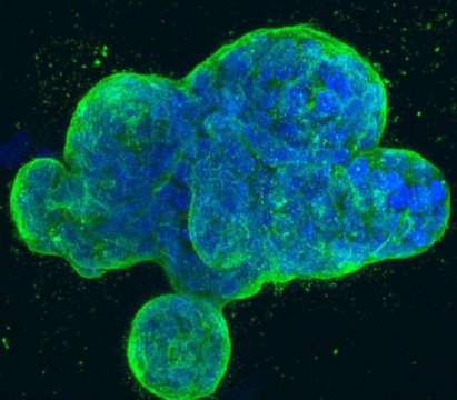
This is a three-dimensional culture of human breast cancer cells, with DNA stained blue and a protein in the cell surface membrane stained green. Credit: National Institutes of Health

Adenosine deaminases are a family of three enzymes encoded by the ADAR genes, which stand for adenosine deaminase acting on RNA. They regulate gene expression by modifying nucleotides within double stranded RNA molecules, serving as fundamental editors in the development of new stem cells.

The enzyme, however, is also activated in cancers as diverse as liver, breast and leukemia. A research team led by senior author Catriona Jamieson, MD, PhD, deputy director of the Sanford Stem Cell Clinical Center and deputy director of the UC San Diego Moores Cancer Center, found that the normal functions of the ADAR1 enzyme are hijacked by pre-malignant cells, leading to a cascade of molecular consequences that promote malignant transformation, dormant cancer stem cell generation and resistance to treatment.
"We were able to illuminate the abilities of ADAR1 to 'hyper-mutate' tumor suppressor RNAs in leukemia and, at the same time, edit the microRNA aimed at targeting the tumor suppressor RNA. This enzyme turns on cancer resistance via a domino effect on RNA instead of DNA," said Qingfei Jiang, PhD, Assistant Project Scientist, Jamieson's lab.
Jamieson characterized RNA editing as tweaking basic genetic blueprints, not fundamentally rewriting them. Nonetheless, the results might be dramatic. "One result of detection of malignant RNA editing could be exposing dormant cancer stem cells that often escape therapies that target dividing cells, which leads to therapeutic resistance and disease relapse, and also highlight ADAR as a potentially tractable target for cancer stem cell elimination," said Jamieson.
Researchers from different part of the world are invited to submit abstract on their unpublished latest research at our upcoming conference Cell Tissue Science 2019 which is focused on the complications and consequences of Stem Cell, Regenerative Medicine, Stem Cell Therapy, Cancer Cell Biology,Technical Advancements in cancer treatment and many more. We as committee members of the conference welcome you to be a part of the conference “ 12th World Congress on Cell & Tissue Science” in Singapore on March 11-12, 2019. 
You can submit your abstract on Session or Track : 09. Stem Cells Biology With Regards to Christmas and New Year Celebration we are providing a special discount of 30% on all Registration Categories for more information please  visit by Click Here

Sunday, 23 December 2018

A compound being developed to treat eye disease also kills leukemia cells


An active ingredient in eye drops that were being developed for the treatment of a form of eye disease has shown promise for treating an aggressive form of blood cancer. Scientists at the Wellcome Sanger Institute, University of Cambridge, University of Nottingham and their collaborators have found that this compound, which targets an essential cancer gene, could kill leukemia cells without harming non-leukemic blood cells.

The results was published in Nature Communications reveal a potential new treatment approach for an aggressive blood cancer with a poor prognosis.

Acute myeloid leukemia (AML) is a form of blood cancer that affects people of all ages, often requiring months of intensive chemotherapy and prolonged hospital admissions. It develops in cells in the bone marrow crowding out the healthy cells, in turn leading to life-threatening infections and bleeding.

Mainstream AML treatments have remained unchanged for over thirty years, with the current treatment being chemotherapy, and the majority of people's cancer cannot be cured. A subtype of AML, driven by rearrangements in the MLL gene has a particularly bad prognosis.

In a previous study, researchers at the Sanger Institute developed an approach, based on CRISPR gene editing technology, which helped them identify more than 400 genes as possible therapeutic targets for different subtypes of AML. One of the genes, SRPK1, was found to be essential for the growth of MLL-rearranged AML. SRPK1 is involved in a process called RNA splicing, which prepares RNA for translation into proteins, the molecules that conduct the majority of normal cellular processes, including growth and proliferation.

In a new study, Sanger Institute researchers and their collaborators set out to work out how inhibition of SRPK1 can kill AML cells and whether it has therapeutic potential in this disease. They first showed that genetic disruption of SRPK1 stopped the growth of MLL-rearranged AML cells and then went on to study the compound SPHINX31, an inhibitor of SRPK1, which was being used to develop an eye drop treatment for retinal neovascular disease -- the growth of new blood vessels on the retinal surface that bleed spontaneously and cause vision loss.

The team found that the compound strongly inhibited the growth of several MLL-rearranged AML cell lines, but did not inhibit the growth of normal blood stem cells. They then transplanted patient-derived human AML cells into immunocompromised mice and treated them with the compound. Strikingly, the growth of AML cells was strongly inhibited and the mice did not show any noticeable side effects.
Dr George Vassiliou, Wellcome Sanger Institute and the Wellcome-MRC Cambridge Stem Cell Institute, said: "We have discovered that inhibiting a key gene with a compound being developed for an eye condition can stop the growth of an aggressive form of acute myeloid leukemia without harming healthy cells. This shows promise as a potential approach for treating this aggressive leukemia in humans."
SRPK1 controls the splicing* of RNA in the production of new proteins. An example of a gene that is affected when SRPK1 is blocked is BRD4, a well-known gene that maintains AML. Inhibiting SRPK1 causes the main form of BRD4 to switch to another form, a change that is detrimental to AML growth.
Dr Konstantinos Tzelepis, the Wellcome Sanger Institute and University of Cambridge, said: "Our study describes a novel mechanism required for leukemia cell survival and highlights the therapeutic potential of SRPK1 inhibition in an aggressive type of AML. Targeting this mechanism may be effective in other cancers where BRD4 and SRPK1 play a role, such as metastatic breast cancer."
Professor David Bates, University of Nottingham and co-founder of biotech company Exonate, which develops eye drops for retinal diseases, said: "When Dr Vassiliou told me that SRPK1 was required for the survival of a form of AML, I immediately wanted to work with him to find out if our inhibitors could actually stop the leukemia cells growing. The fact that the compound worked so effectively bodes well for its potential development as a new therapy for leukemia. It will take some time, but there is real promise for a new treatment on the horizon for patients with this aggressive cancer."
We welcome researchers from different part of the to submit abstract on their latest research at our upcoming conference Cell Tissue Science 2019 which is mainly focuses on the complications the consequences of Stem Cell, Regenerative Medicine, Stem Cell Therapy, Cancer Cell Biology,Technical Advancements in cancer treatment and many more.We welcome you to the our upcoming conference “ 12th World Congress on Cell & Tissue Science” . 
You can submit your abstract on Session or Track : 09. Stem Cells and its Applications

Tuesday, 18 December 2018

Depression, anxiety may take same toll on health as smoking and obesity


An annual physical typically involves a weight check and questions about unhealthy habits like smoking, but a new study from UC San Francisco suggests health care providers may be overlooking a critical question: Are you depressed or anxious?

Anxiety and depression may be leading predictors of conditions ranging from heart disease and high blood pressure to arthritis, headaches, back pain and stomach upset, having similar effects as long-established risk factors like smoking and obesity, according to the new research.
In the study looked at the health data of more than 15,000 older adults over a four-year period.
They found that 16 percent (2,225) suffered from high levels of anxiety and depression, 31 percent (4,737) were obese and 14 percent (2,125) were current smokers, according to their study published in the journal Health Psychology on Dec. 17, 2018.

Participants with high levels of anxiety and depression were found to face 65 percent increased odds for a heart condition, 64 percent for stroke, 50 percent for high blood pressure and 87 for arthritis, compared to those without anxiety and depression.
"These increased odds are similar to those of participants who are smokers or are obese," said O'Donovan, who, with Niles, also is affiliated with UCSF Weill Institute for Neurosciences. "However, for arthritis, high anxiety and depression seem to confer higher risks than smoking and obesity."
Cancer an Exception to Conditions Impacted by Depression and Anxiety
Unlike the other conditions investigated, the authors found that high levels of depression and anxiety were not associated with cancer incidence. This confirms results from previous studies, but contradicts a prevailing idea shared by many patients.
"Our findings are in line with a lot of other studies showing that psychological distress is not a strong predictor of many types of cancer," O'Donovan said. "On top of highlighting that mental health matters for a whole host of medical illnesses, it is important that we promote these null findings. We need to stop attributing cancer diagnoses to histories of stress, depression and anxiety."
Niles and O'Donovan discovered that symptoms such as headache, stomach upset, back pain and shortness of breath increased exponentially in association with high stress and depression. Odds for headache, for example, were 161 percent higher in this group, compared with no increase among the participants who were obese and smokers.
Treating Mental Health Can Cut Health Care Costs
"Anxiety and depression symptoms are strongly linked to poor physical health, yet these conditions continue to receive limited attention in primary care settings, compared to smoking and obesity," Niles said. "To our knowledge this is the first study that directly compared anxiety and depression to obesity and smoking as prospective risk factors for disease onset in long-term studies."
The results of the study underscore the "long-term costs of untreated depression and anxiety," said O'Donovan. "They serve as a reminder that treating mental health conditions can save money for health systems."
The two authors evaluated health data from a government study of 15,418 retirees, whose average age was 68. Depression and anxiety symptoms were assessed using data from participant interviews. Participants were questioned about their current smoking status, while weight was self-reported or measured during in-person visits. Medical diagnoses and somatic symptoms were reported by participants.
We welcome researchers from different part of the to submit abstract on their latest research at our upcoming conference Cell Tissue Science 2019 which is mainly focuses on the complications the consequences of Stem Cell, Regenerative Medicine, Stem Cell Therapy, Cancer Cell Biology , Technical Advancements in cancer treatment and many more.We welcome you to the our upcoming conference “ 12th World Congress on Cell & Tissue Science” . 
You can submit your abstract on Session or Track : 08.Cancer Cell Biology

Saturday, 15 December 2018

The source of stem cells points to two proteins

New MSU research published in the journal eLife has pinpointed two proteins that are the keys to stem cell creation.
Credit: Courtesy of MSU
Mammalian embryos are unlike those of any other organism as they must grow within the mother's body. While other animal embryos grow outside the mother, their embryonic cells can get right to work accepting assignments, such as head, tail or vital organ. By contrast, mammalian embryos must first choose between forming the placenta or creating the baby.

New research at Michigan State University and published in the journal eLife has pinpointed two proteins that are the keys to this decision making. The process of assigning cells to placenta or baby is important because that is when pluripotent cells are made. These adaptable pluripotent cells are critical to stem cell research, and these two proteins could be the key to deciphering how pluripotent cells are born, said Amy Ralston, MSU's inaugural James K. Billman Jr., M.D. endowed professor and the study's senior author.
"Pluripotent cells are the progenitors of embryonic stem cells, and they are famous because they can become any part of the body," she said. "We have discovered a process that regulates the balance between pluripotent and placenta, and it works by changing the physical location of cells within the embryo's ball of cells."
To form this wondrous ball, the mother packages two closely related proteins into her eggs, which help oversee this decision-making process. The scientific team used genetic tools to eliminate the proteins YAP1 and WWTR1 from mouse eggs and embryos. The team discovered that these two proteins first position these cells into distinct inside and outside locations, which then decides their fate: placenta or pluripotent.

Each phase of Ralston's research peels back a layer into the creation of stem cells, a process that nature performs with 100 percent efficiency. On the other end of the spectrum, lab-created stem cells are created with 1 percent efficiency.
"Obviously, our understanding of how nature creates stem cells is incomplete," said Tristan Frum, MSU biochemist and molecular biologist"We've suspected these proteins were involved in creating stem cells, but our work reveals that they do so in a surprising way."
Cells of the early embryo express proteins that make them "stick" to the outside of the embryo.
"We've identified a way that cells evade this stickiness, crawl inside the embryo and acquire the properties that make stem cells so interesting and useful," Frum said. "When we make stem cells in the lab, they must acquire a unique cell membrane. Our work shows how nature does this and provides clues that can guide us to control stem cells more efficiently for use in medicine".
For example, the team's discoveries could lead to advances in stem cell technologies using organoids, which are stem cell-derived mini organs. Organoidsare an exciting new paradigm in stem cell and regenerative medicine, The similarities between embryos and organoids are remarkable. Therefore, by studying how the mouse embryo builds itself, we may one day build organs from human stem cells." says Ralston.
We welcome researchers from different part of the to submit abstract on their latest research at our upcoming conference Cell Tissue Science 2019 which is mainly focuses on the complications the consequences of Stem Cell, Regenerative Medicine, Stem Cell Therapy, Cancer Cell Biology,Technical Advancements in cancer treatmentand many more.We welcome you to the our upcoming conference “ 12th World Congress on Cell & Tissue Science” . 
You can submit your abstract on Session or Track : 09. Stem Cells and its Applications

Friday, 14 December 2018

Excess body weight responsible for nearly 4 percent of cancers worldwide


Policies, economic systems, and marketing practices that promote the consumption of energy-dense, nutrient-poor food, changing behavioral patterns that couple high total energy intake with insufficient physical activity, and human-built environments that amplify these factors are driving a worldwide rise in excess body weight, according to a new report. The report, appearing early online in CA: A Cancer Journal for Clinicians, a peer-reviewed journal of the American Cancer Society, says excess body weight accounted for approximately 3.9% of all cancers worldwide in 2012, a figure that will undoubtedly rise in the coming decades given current trends.

The prevalence of excess body weight has been increasing worldwide since the 1970s. In 2016, approximately 40% of adults and 18% of children (ages 5-19 years) had excess body weight, equating to almost 2 billion adults and 340 million children around the globe. The report says the prevalence of excess body weight has increased rapidly in most countries across all population groups. Some of the steepest increases are in low- and middle-income countries, likely the result of the spread of the "Western lifestyle," consisting of energy-dense, nutrient-poor foods alongside reduced physical activity levels.

In 2015, an estimated 4 million deaths were attributable to excess body weight. The worldwide economic impact of illness related to excess body weight is estimated at $2.0 trillion. In 2012, excess body weight accounted for approximately 544,300 cancers, 3.9% of all cancers worldwide, with the proportion varying from less than 1% in low-income countries to 7% or 8% in some high-income Western countries and in Middle Eastern and Northern African countries.

Overweight and obesity has been linked to an increased risk of 13 cancers: cancers of the breast (postmenopausal), colon and rectum (colorectal), corpus uteri, esophagus (adenocarcinoma), gallbladder, kidney, liver, ovary, pancreas, stomach (cardia), and thyroid, as well as meningioma and multiple myeloma. More recently, overweight has been labeled a probable cause of advanced prostate cancer as well as cancers of the mouth, pharynx, and larynx.

National wealth is the most apparent systematic driver of population obesity. The economic transition to a wealthier economy brings with it an environment that precipitate obesity. Each $10,000 increase in average national income is associated with a 0.4 increase in body mass index among adults. However, prosperity is not always correlated with excess body weight; obesity prevalence is quite low in high-income Asian Pacific countries (range, 4%-7%), which is likely a result of adherence to traditional dietary habits, which are conducive to lower calorie consumption, and an active transportation system that usually entails walking as part of daily activity. Meanwhile, the prevalence of obesity is very high in some lower-income countries, such as some Pacific Island nations (range, 40%-65%) and Egypt (43% among women and 24% among men).

Halting the rise in obesity is one of the World Health Organization (WHO)'s nine 2025 targets to address the growing global burden of noncommunicable diseases, including cancer. While the current pace of increasing and existing challenges makes achieving this goal appears unlikely, the WHO says high-priority strategies that should be adopted by governments, industries, and civil societies include population-wide, policy-led interventions to rectify the production, distribution, and marketing of unhealthy foods and changes in the built environment to promote adequate levels of physical activity.

Those interventions include eliminating trans-fats through the development of legislation to ban their use in the food chain; reducing sugar consumption through effective taxation on sugar-sweetened beverages; implementing subsidies to increase the intake of fruits and vegetables; limiting portion and package size to reduce energy intake and the risk of excess body weight; ensuring that urban design incorporates the core elements of residential density, connected street networks that include sidewalks, easy access to a diversity of destinations, and access to public transport; and providing convenient and safe access to quality public open space and adequate infrastructure to support walking and cycling.
"There is emerging consensus on opportunities for obesity control through the multisectoral coordinated implementation of core policy actions to promote an environment conducive to a healthy diet and active living," says Hyuna Sung, American Cancer Society scientist."The rapid increase in both the prevalence of excess body weight and the associated cancer burden highlights the need for a rejuvenated focus on identifying, implementing, and evaluating interventions to prevent and control excess body weight."
We welcome researchers from different part of the to submit abstract on their latest research at our upcoming conference Cell Tissue Science 2019 which is mainly focuses on the complications the consequences of Stem Cell, Regenerative Medicine, Stem Cell Therapy, Cancer Cell Biology , Technical Advancements in cancer treatment and many more.We welcome you to the our upcoming conference “ 12th World Congress on Cell & Tissue Science” .  You can submit your abstract on Session or Track : 08.Cancer Cell Biology
   

Thursday, 13 December 2018

Unlocking the secrets of how cells communicate offers insights into treating diseases


2D images, left, and the 3D structure, right, of the gap junction communication channel from the eye lens, resolved to near-atomic resolution by the Reichow Lab using cryo-electron microscopy.
Credit: Reichow Lab | Portland State University
Portland State University researchers have made a significant breakthrough by developing the 3-D structure of proteins from inside the eye lens that control how cells communicate with each other, which could open the door to treating diseases such as cataracts, stroke and cancer.

The PSU research team, led by chemistry professor Steve Reichow, used a multimillion-dollar microscope and a novel technique developed by three Nobel Prize-winning biophysicists called cryo-electron microscopy (Cryo-EM) to view membrane protein channels or transportation tunnels in cell walls -- at the atomic level. This allowed Reichow's team, whose research is supported by the National Institutes of Health, to create a 3-D image of the membrane channel to better understand the processes involved in cell-to-cell communication.

Portland State researchers used Cryo-EM a microscope technique that freezes biomolecules in mid-movement and takes ultra-high-resolution images and computer modeling to see the 3-D structure of gap junction proteins that had been isolated from eye lenses. Gap junctions are tiny channels that allow neighboring cells to communicate with one another and are found in many places throughout the body.

Their findings published in the scientific Journal Nature -- showed for the first time how gap junctions selectively pass or block chemical information. Until now, it was not known how these channels would allow certain messages to pass between cells while specifically blocking others.

Reichow said the detailed images may open the door to understanding, and potentially treating, different types of diseases that are associated with the loss of function in cellular communication involving gap junctions such as cataracts, cardiac arrhythmia, stroke and certain cancers.
"Currently there's no drug on the market today that can specifically block or activate gap junction proteins," Reichow said. "But our discovery may one day pave the way for development of pharmaceuticals that can control heart disease or other diseases that are associated with the malfunctions or mis-regulations of these protein channels."
Now that the Reichow Lab has established a method for characterizing the proteins, they are trying to understand the nuances of how the body uses gap junctions differently across tissues and organs.
We welcome researchers from different part of the to submit abstract on their latest research at our upcoming conference Cell Tissue Science 2019 which is mainly focuses on the complications the consequences of Stem Cell, Regenerative Medicine, Stem Cell Therapy, Cancer Cell Biology , Technical Advancements in cancer treatment and many more.We welcome you to the our upcoming conference “ 12th World Congress on Cell & Tissue Science” . 
You can submit your abstract on Session or Track : 09. Stem Cells and its Applications

Tuesday, 11 December 2018

Potential seen for tailoring treatment for acute myeloid leukemia


Advances in rapid screening of leukemia cells for drug susceptibility and resistance are bringing scientists closer to patient-tailored treatment for acute myeloid leukemia (AML).

Research on the drug responses of leukemia stem cells may reveal why some attempts to treat are not successful or why initially promising treatment results are not sustained.

AML is a serious disorder of certain blood-forming cells. In this disease, certain early precursor cells in the bone marrow that usually develop into white blood cells don't mature properly. They remain frozen as primitive cells called blasts, unable to further differentiate and mature. These can accumulate and cause low blood counts that reduce the ability to fight infections, and low platelet counts that cause risk of life threatening hemorrhage.

Leukemia stem cells -- the progenitors for the immature, cancerous blood cells -- propagate AML, and also play a role in the cancer returning after treatment. Cancer researchers are interested in how genes are expressed in this cell population, because this data may hold clues to resistance to standard therapies and answers to why some patients relapse.

A study presented at the 60th Annual Meeting of the American Society of Hematology in San Diego looked at the drug response patterns of stem cells and blast cells taken from individual patients diagnosed with acute myeloid leukemia. The information was gathered through high throughput screening, a state of the art method for quickly evaluating and testing many samples.

The researchers found that leukemia stem cells and blast cells diverged in their drug susceptibility patterns, and also that these patterns differed from patient to patient.
For example: Blast cells s responded in the test to the drugs most commonly used to treat patients, but none were effective against leukemia stem cells. The researchers did find 12 drugs from eight classes that seemed to preferentially target leukemia stem cells, compared to blast cells. Many of them are not often used in patients with this type of cancer.
The multidisciplinary team on the project included stem cell biologists, hematologists, medical oncologists, pathologists, computer scientists, drug developers and others.

The senior researcher was hematologist Dr. Pamela Becker, professor of medicine at the UW School of Medicine. She is also a scientist at the Fred Hutchinson Cancer Research Center and the UW Medicine Institute for Stem Cells and Regenerative Medicine, and sees patients with blood disorders at the Seattle Cancer Care Alliance.

In the laboratory study, the researchers compared the drug sensitivity of blast cell and stem cell populations taken from the same six patients. In doing so, they tested a custom panel of drugs, targeted agents and drug combinations on the cells, and did genetic analyses for 194 mutations. The panel included both FDA approved and investigational drugs.

The unique drug susceptibility patterns observed in leukemia stem cells and blast cells are leading the scientists to hope that patient-specific approaches could be developed against acute myeloid leukemia, with the goal of improving the outcomes for people with this form of blood cancer.
We welcome researchers from different part of the to submit abstract on their latest research at our upcoming conference Cell Tissue Science 2019 which is mainly focuses on the complications the consequences of Stem Cell, Regenerative Medicine, Stem Cell Therapy, Cancer Cell Biology , Technical Advancements in cancer treatmentand many more.We welcome you to the our upcoming conference “ 12th World Congress on Cell & Tissue Science” . 
You can submit your abstract on Session or Track : 09. Stem Cells and its Applications

Friday, 7 December 2018

Platelets grown from stem cells may be alternative to donated platelets


Researchers have developed a way to grow human platelets in the laboratory from stem cells derived from fat tissue. The achievement, reported today in the Journal Blood, suggests manufactured platelets could eventually reduce the reliance on donated platelets to help patients with cancer and other disorders.

Platelets are a component of blood that helps with clotting. Platelet transfusions can be life-saving for patients dealing with cancer or the effects of chemotherapy, infections, immune disorders, or platelet disorders.

Over 4.5 million platelet units of plasma are transfused every year worldwide, a need that currently must be met by human donors. Because donated platelets have a shelf life of less than a week, supplies often fall short of patient needs. In addition, donated platelets are subject to inherent safety risks due to infection from the donor and immune response in the recipient.
"By removing the donor from the equation, adipose-derived stem cells could be used to provide a ready supply of safe, tolerable platelets to meet an ever-changing demand," said Dr. Mastubara.
Researchers initially sought to derive platelets from two other types of stem cells including one known as induced pluripotent stem cells (iPS). They noticed adipose-derived cells, which were being used as a negative control in the experiments, had produced megakaryocyte- and platelet-sized cells that naturally expressed several genes important to producing platelets.

After refining methods for coaxing adipose-derived stem cells to produce platelets, the researchers conducted a series of tests to determine whether the manufactured platelets would function similarly to natural human platelets. They verified that the lab-grown platelets contained hallmark proteins found on the surface of natural platelets, as well as granules that are key to the clotting process. Blood clotting simulations and experiments using mice confirmed that the platelets behave like donated platelets, gathering together into clumps to form clots.
"Though more expensive to harvest compared to donor-derived platelets, this research demonstrates that platelets can be produced from adipose-derived cells by a rather simple method," said Matsubara. "Now that we have established an efficient manufacturing process to yield a large number of adipose-derived platelets, we next plan to perform preclinical studies using animal models to demonstrate efficacy and safety, followed by clinical trials in human patients."
We welcome researchers from different part of the to submit abstract on their latest research at our upcoming conference Cell Tissue Science 2019 which is mainly focuses on the complications the consequences of Stem Cell, Regenerative Medicine, Stem Cell Therapy, Cancer Cell Biology , Technical Advancements in cancer treatmentand many more.We welcome you to the our upcoming conference “ 12th World Congress on Cell & Tissue Science” .  You can submit your abstract on Session or Track : 09. Stem Cells and its Applications

Thursday, 6 December 2018

Citrate-based biomaterial fuels bone healing with less rejection


A material based on a natural product of bones and citrus fruit, called citrate, provides the extra energy that stem cells need to form new bone tissue, according to a team of Penn State bioengineers. Their new understanding of the mechanism that allows citrate to aid in bone regeneration will help the researchers develop slow-release, biodegradable, citrate-releasing scaffolds to act as bone-growth templates to speed up healing in the body.
"In our lab, we have been working with citrate for over a decade," said Jian Yang, Professor of Biomedical Engineering, Penn State. "We knew that in the human body, 90 percent of organic citrate is located in skeletal tissue. But no one had really tried to use citrate as a building block to make bone biomaterials. Our new paper tries to understand how citrate helps in bone healing and uses the understanding to guide the design of new biomimetic biomaterials for better bone repair."
Autografting -- taking bone from another part of the patient's body and grafting it to the wound -- is the method used most for bone regeneration in a hospital setting. This is not always a suitable method, especially in the case of large wounds or bone tissue removed during cancer treatment.

Synthetic biomaterials would be a welcome replacement and many labs are working on developing them. But current synthetic materials cause significant inflammation, and the bone healing rate is slow and healing quality is poor. The body encapsulates the implant with fibrotic tissues that keep the implant from integrating with surrounding bone. With Yang's material, the researchers do not see encapsulation, and chronic inflammation is minimal.

Chuying Ma, a doctoral student in Yang's lab, is lead author on the paper. Ma was given the problem of uncovering the poorly known mechanism underlying the body's use of citrate to regenerate bone. She found that the stem cell membrane has a transporter that is used to transport citrate into the cell to elevate the cellular energy level. When the bone stem cells differentiate to make new bone cells, they require more energy as support for active bone formation. The timing and dosage of citrate supply to stem cells are also critical. Yang and Ma coined the newly identified citrate effect on stem cell differentiation as "metabonegenic regulation" in the paper.

The team also identified a second factor involved in energy production, an amino acid called phosphoserine. With their new understanding of the mechanism for bone regrowth, they developed a biomaterial incorporating both citrate and phosphoserine and tested it on rat models.
"Using our new material, we see the early deposition of new bone at one month," Ma said. "This is much earlier than the biomaterials widely used in FDA-approved devices. In this study we tested two models, the femoral condyle bone and cranial bone defects."
In both animal models we see the new biomaterial is better than the commercial materials in inducing early bone formation and also promoting bone maturation, according to Ma. "To me, this is an important finding," said Yang, who is a faculty member in Penn State's Materials Research Institute and the Huck Institutes of the Life Sciences. "Citrate is now recognized as a central linker between stem cell metabolism and differentiation. We are uncovering the mechanism whereby citrate influences stem cell activity, not only in bone, but by implication extending to other types of cells and tissues. For instance, there is a high concentration of citrate in the cerebrospinal fluid surrounding the brain. People can now use this understanding to start looking at citrate as a metabolism regulator to further regulate stem cells for other types of tissues and organs throughout the body."
We welcome researchers from different part of the to submit abstract on their latest research at our upcoming conference Cell Tissue Science 2019 which is mainly focuses on the complications the consequences of Stem Cell, Regenerative Medicine, Stem Cell Therapy, Cancer Cell Biology , Technical Advancements in cancer treatment and many more.We welcome you to the our upcoming conference “ 12th World Congress on Cell & Tissue Science” . 
You can submit your abstract on Session or Track : 10.Biomaterial and Bioengineering