Showing posts with label Top Stem Cell Conferences. Show all posts

Saturday, 19 January 2019

How stem cells self-organize in the developing embryo


Embryonic development is a process of profound physical transformation, one that has challenged researchers for centuries. How do genes and molecules control forces and tissue stiffness to orchestrate the emergence of form in the developing embryo? How are the precise mechanics underlying emergence of the complexity of our organs and tissues encoded in our DNA?

One particular aspect of embryonic development: how a group of stem cells -- the endoderm -- moves from the surface of the developing embryo to the center, and in doing so transforms from a flat sheet to a hollow tube. This structure, known as the gut tube, then forms the lining of the entire respiratory and gastrointestinal tracts.

In a study published today in Nature, Nerurkar worked with colleagues at Harvard to shed new light on this critical step in early embryonic development. The team discovered gut tube formation is driven by collective cell movements of the endoderm, a process by which cells travel large distances en masse, without rearranging relative to one another. They also found that this collective movement is triggered by cells that are converting a molecular gradient to a force gradient that drives cells from the surface into the embryo. This discovery is one of the few examples, especially among vertebrates, of how molecular cues are converted into the physical forces that shape our organs.
The study findings could have important implications for how stem cells are used to create functional organs in the lab, and lead to a better understanding of the underlying causes of gastrointestinal birth defects. "Our major goal is to understand how we, as complex organisms, are formed with such precision from a seemingly disorganized ball of cells -- the early embryo," says Nandan Nerurkar, assistant professor of biomedical engineering at Columbia Engineering.
Identifying genes that drive differentiation of stem cells into mature cell types -- the primary focus in Nerurkar's field -- is an important step toward growing replacement organs in the lab. However, Nerurkar suggests this is only part of the picture: "It is equally important to understand how to instruct those cells to organize into functional three-dimensional organs. The developing embryo holds the recipe for this, and many research groups, including ours, are now leveraging the language of physics and mechanics to dissect it."

The team used an innovative approach at the leading edge of the developmental biology field. They combined conventional approaches of developmental biology, including analysis and manipulation of gene expression and live time lapse microscopy of cell movements in the developing chick embryo, with engineering methods, such as mathematical modeling and force and strain measurements.

They focused on one part of endoderm internalization: the hindgut, which gives rise to half of the small intestine, the large intestine, and colon. What was previously known of gut tube formation came from fate-mapping experiments, wherein cells are labeled early in development and then mapped to where the labeled cells end up later in development. This static analysis, which uses static images of the beginning and the end of the process to make an educated guess of what happens in the middle, has led to a view of gut tube formation that is present in most embryology textbooks. "Based on our recent findings, this view is at best incomplete, and at worst completely wrong," says Nerurkar.

Unlike earlier fate-mapping studies, Nerurkar and his colleagues used live imaging in the embryo to directly observe cell movements as the endoderm is internalized to form a tube. They next applied a combination of mechanical engineering and developmental biology approaches to understand just how those cell movements occur, and how the movements are coordinated to form this critical structure in the early embryo.

The team found that the movements are coordinated by the conversion of a molecular gradient into a force gradient from cells that are contracting in proportion to the amount of a molecular cue -- fibroblast growth factor (FGF) -- that they sense. This results in a tug of war among endoderm cells: as one "team" begins to win, the cells actually recruit players from the opposing team by pulling them from low to higher concentrations of FGF.
Irregularities in FGF function can lead to a number of developmental defects. "During human development, errors in gut tube formation would likely lead to miscarriage, something that is a relatively high risk during the first trimester, when this process is occurring," says Nerurkar.
While this study focused on just one part of endoderm internalization, the hindgut, it is still unknown how the foregut, which forms the trachea, lungs, esophagus, stomach, and liver, and the midgut, which forms the pancreas and small intestine, are formed. Nerurkar plans to use his new approach to study these other areas of embryonic development and investigate if and how FGF signaling acts more broadly to control mechanics in the development of other tissues and organs.
"I want to learn more about how mechanics and molecules are integrated to coordinate the formation of these very distinct tissues by disparate mechanisms, yet from the same initial pool of stem cells," he says. "By focusing on the tissue-level mechanics downstream of FGF signaling, we may now be able to understand what this important pathway does to shape other organs and tissues during development, including the heart, brain, and spinal column."
Nerurkar is continuing this research at Columbia Engineering, developing quantitative molecular-mechanical relationships that could be used to design and construct replacement tissues in the lab, using controlled delivery of these diffusible cues -- the instructional signals that are secreted by cells and then float away to neighboring cells -- to instruct the self organization of cells into functional tissues and organs. If he and others in this field can establish the design principles of embryonic tissue formation, it will be possible to repurpose those same principles for regenerative medicine and tissue engineering applications.
Researchers from different part of the world are invited to submit abstract on their unpublished latest research at our upcoming conference Cell Tissue Science 2019 which is focused on the complications and consequences of Stem CellRegenerative MedicineStem Cell TherapyCancer Cell Biology,Technical Advancements in cancer treatment and many more. We as committee members of the conference welcome you to be a part of the conference “ 12th World Congress on Cell & Tissue Science” in Singapore on March 11-12, 2019
You can submit your abstract on Session or Track : 09. Stem Cells and its Applications

Friday, 18 January 2019

Mathematical model can improve our knowledge on cancer


Researchers have developed a new mathematical tool, which can improve our understanding of what happens when cells lose their polarity (direction) in diseases such as cancer. The result is advancing our understanding of how the fertilized egg cell develops into a complete organism. Biological shapes, like individual organs or an entire body, can be reproduced or maintained with great accuracy, just like in the embryonic development or during the adult stage.

It remains unknown how cells "know" which structures to form in order to repair tissue damage:

Multicellular organisms can develop highly complex structures that make up their tissue or organs and are capable of regenerating perfect reproductions of these structures after injury. This involves folding of sheets, formed by groups of dividing and interacting cells. Yet, although much is understood about some of the intermediate steps that occur during development and tissue repair, exactly how thousands of cells together work out what shapes they need to form remains unknown.
Building the mathematical model:
"In this study, we wanted to see how cells organize into folded sheets and tubes, and how this process can be so precisely reproduced as is seen during development," says lead author Silas Boye Nissen, PhD student at the Center for Stem Cell Decision Making, StemPhys, University of Copenhagen, Denmark. "To answer this question, we built a mathematical tool that can model two types of cell polarities and simulated how many cells organize themselves into folded sheets and organs."
The researchers found that by changing one of two polarities in the model, they were able to simulate a rich diversity of shapes. The differences in the shapes were dictated by two factors: The initial arrangement of the cells and external boundaries -- such as the shape of an egg influencing the development of the embryo inside.

By exploring a multitude of theoretical scenarios in which the polarities were altered, the model was able to narrow down the focus to a few theories to be tested experimentally. In miniaturized versions of organs grown in the lab (called organoids), the model predicted that rapid, off-balance growth of cells will cause the growing organoid to develop lots of shallow folds, while external pressure caused by the medium on the organoids will cause fewer, deeper and longer folds. This means the model can improve our understanding of how folded organs like the brain or the pancreas are formed.
Few, simple rules apply for the formation of biological shapes:
"Our findings advance our understanding of how properties of individual cells lead to differences in shapes formed by thousands of cells," says senior author Professor Kim Sneppen, Director of the Center for Models of Life, CMOL, University of Copenhagen, and senior coauthor Ala Trusina concludes: "Our work suggests that body parts may not need detailed instructions to form, but instead can emerge as cells follow a few simple rules. We can now explore what happens if cells gain or lose their polarities at the wrong time or place, as often happens in cancer."
Researchers from different part of the world are invited to submit abstract on their unpublished latest research at our upcoming conference Cell Tissue Science 2019 which is focused on the complications and consequences of Stem CellRegenerative MedicineStem Cell Therapy, Cancer Cell Biology,Technical Advancements in cancer treatment and many more. We as committee members of the conference welcome you to be a part of the conference “ 12th World Congress on Cell & Tissue Science” in Singapore on March 11-12, 2019
You can submit your abstract on Session or Track : 08. Advancement in Cancer Treatments

Monday, 26 November 2018

Underlying mechanisms of 3d tissue formation


If you want to build an organ, such as for transplant, you need to think in 3D.
Using stem cells, scientists for some time have been able to grow parts of organs in the lab, but that is a far cry from constructing an actual, fully-formed, functioning, three-dimensional organ.

For students of regenerative medicine and developmental biology, this is why understanding how cells bend and move to form organs and bodily tissue is a hot topic.

And now a team at Kyoto University's Institute for Frontier Life and Medical Sciences have gained new understanding into how cells undergoing mechanical strain create the spherical structure of the eye.

Publishing in Science Advances, the team has found that individual cells together form a primordial, cup-like structure -- an 'optic cup' -- by sensing mechanical forces resulting from the deformation of the entire tissue.
"In the past, we succeeded in making the optic cup by culturing embryonic stem -- ES -- cells. To form a sphere, the tissue needed to first protrude from primordial brain tissue and then invaginate inside," explains first author Satoru Okuda."But how individual cells sensed and modulated themselves to form that shape had been unclear."
The team developed a computational simulation that calculates the formation of three-dimensional tissue structures. Using this knowledge and past experimental data, they constructed a virtual precursor-eye and were able to predict the physics driving the sphere-forming cells.

Their findings show that during optic cup formation, a cell differentiation pattern -- pushing cells into the cup shape -- is generated, causing a portion of the cells to spontaneously fold into the tissue. This force caused by 'self-bending' propagates to the boundary region, where other cells sense the strain.
"The combination of the tissue deformation and the strain on the boundary of the optic cup generates a hinge that further pushes the bending cells," continues Okuda, "leading to the cup-like structure.The next step was to verify this prediction using actual ES cells."
Utilizing mouse ES cells in culture, the team applied mechanical strain on specific points and were pleased to detect the calcium responses, mechanical feedback, and cell shape changes they had predicted in the simulations.

These findings reveal a new role for mechanical forces in shaping organs, which is crucial in forming complex tissues, even in a petri dish. The team will continue to investigate these forces, seeking to continue advancing the field of regenerative medicine.
"While our research shows the possibility of controlling the shapes of organs made in vitro -- using appropriate mechanical stimulation based on prediction -- current techniques are still limited," concludes lead scientist Mototsugu Eiraku."We hope to improve the predictive accuracy of our simulations and recreate more complicated tissues and organs in the future." 
We welcome researchers from different part of the to submit abstract on their latest research at our upcoming conference Cell Tissue Science 2019 which is mainly focuses on the complications the consequences of Stem CellRegenerative MedicineStem Cell TherapyCancer Cell Biology , Technical Advancements in cancer treatment and many more.We welcome you to the our upcoming conference “ 12th World Congress on Cell & Tissue Science” . 
You can submit your abstract on Session or Track : 15. Regenerative Medicine

Thursday, 15 November 2018

Mitochondrial DNA may have strong influence on cellular metabolism and disease susceptibility

About 1.5 billion years ago, tiny visitors came to live inside the cells that later evolved into all plant and animal life including humans.

Those visitors were mitochondria, small organelles whose prominent role is producing 90 percent of the chemical energy cells need to survive. Evolutionarily speaking, humans, animals and plants are thus a combination of two organisms.

Mitochondria have their own DNA, but the 13 genes in human mitochondria along with DNA sequences for tRNAs, rRNAs and some small peptides are massively overshadowed by the 20,000 genes in the human nucleus. Nevertheless, these diminutive mitochondria may have a strong influence on cellular metabolism and susceptibility to metabolic diseases like heart failure or obesity, according to preliminary research by Scott Ballinger, Ph.D., professor of pathology at the University of Alabama at Birmingham.
"For 50 years, researchers have tried to find disease susceptibility using Mendelian genetics," Ballinger said while speaking about studies of the chromosomal genes in the cell nucleus. "But this explains only 10 percent of the reasons for susceptibility to disease."
The possible impact of mitochondrial DNA on disease susceptibility depends on two facts. First, all of a person's mitochondrial DNA comes from the mother, via her egg. This is distinct from the chromosomal genes in the nucleus, where, on average, half come from the mother and half from the father. Second, human mitochondrial DNA has evolved into distinct haplotypes, and each of these types has mitochondrial DNA variations that are inherited together. There are approximately 25 to 35 basic mitochondrial DNA haplogroups, and one of them is found in African populations and has many subtypes due the deep genetic diversity of that continent.

To investigate the impact of mitochondrial DNA, Ballinger and colleagues looked for changes in metabolism and nuclear gene expression when they exchanged mitochondrial backgrounds of strains of mice -- specifically those having different mitochondrial DNA sequences, and also having notable differences in susceptibility to diseases associated with metabolism.

In the first report of its kind, they found that switching the mitochondrial genetic background had a significant impact on adiposity, whole body metabolism and nuclear gene expression in mice.

For example, gene expression in both visceral and subcutaneous fat was markedly changed between mice sharing the same nuclear genome but having different mitochondrial DNA backgrounds, when fed chow versus a high-fat diet. These changes ranged from 10- to 50-fold differences in the number of genes affected, and mitochondrial DNA background influenced whether the number of affected genes were increased or decreased. These studies also found that metabolic efficiency and percentage of body fat in the mice were impacted as well.
"These results are clearly consistent with the notion that different nuclear-mitochondrial genetic combinations influence metabolism, adiposity and gene expression in different ways," Ballinger said. "The overall implication of this work is that it can provide a new framework for understanding complex genetic disease susceptibility that both an individual's nuclear and mitochondrial genomes, in combination, can affect disease development. We are now trying to understand how the different combinations of nuclear and mitochondrial encoded genes interact to alter metabolism, and how this influences individual disease susceptibility."
We welcome researchers from different part of the to submit abstract on their latest research at our upcoming conference Cell Tissue Science 2019 which is mainly focuses on the complications the consequences of Stem CellRegenerative MedicineStem Cell TherapyCancer Cell Biology , Technical Advancements in cancer treatment and many more.We welcome you to the our upcoming conference “ 12th World Congress on Cell & Tissue Science” . 
You can submit your abstract on Session or Track :12.Epigenetics and Epigenome

Monday, 8 October 2018

Novel mechanism for generating our skeleton

There are more than 200 bones in the human body. Bone is formed during embryonic and postnatal skeletogenesis by two distinct, well-organized processes, intramembranous and endochondral ossification.

Mesenchymal stem cells differentiate into chondrocytes to form a cartilaginous template, which, for long bones, induces bone formation through endochondral ossification. Extracellular signal-regulated kinase 5 (Erk5), which is a member of the mitogen-activated protein kinase (MAPK) family, is phosphorylated by MAPK/Erk kinase-5 (Mek5) to regulate the function of various cell types.

Although the MAPK Erk1/2 pathway is well known for regulating skeletogenesis, the in vivo physiological role of the Mek5/Erk5 pathway in skeletal development has been largely unclear to date, because of the early embryonic lethality of global Erk5 knockout mice.

181002103035_1_900x600
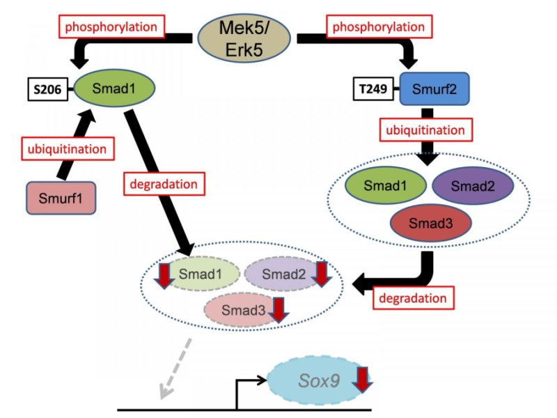
Schematic model of this study.


By using cell-specific mouse genetics approaches revealed that Erk5 plays a crucial role in skeletogenesis in vivo. Paired-related homeobox 1 (Prx1) is expressed in mesenchymal stem cells in the limb buds. Mesenchymal stem cell-specific Erk5 knockout embryos (Prx1-Cre;Erk5fl/fl embryos) displayed wider long bones and impaired mineralization of the metatarsal. In contrast, chondrocyte-specific Erk5 knockout embryos (Col2a1-Cre;Erk5fl/fl embryos) recapitulated only the wider long bone phenotype of Prx1-Cre;Erk5fl/fl embryos. Accordingly, the investigators revealed that Erk5 controls.
  1. Early chondrogenic differentiation of mesenchymal stem cells, including mesenchymal condensation, through its expression in skeletogenic progenitors
  2. Chondrogenic differentiation after the formation of mesenchymal condensations through its expression in chondrocytes.
Subsequent biochemical analyses found that Erk5 directly phosphorylates Smad1 at Ser206 in the linker region, which is known to trigger its proteasomal degradation in a Smad-specific E3 ubiquitin ligase 1 (Smurf1)-dependent manner. In addition, Erk5 directly phosphorylates Smurf2 at Thr249, leading to accelerated proteasomal degradation of Smad proteins (Smad1, 2 and 3).

The study demonstrated that Smads transcriptionally activate the expression in mesenchymal stem cells of sex-determining region within the Y-type high-mobility group box protein 9 (Sox9), which is the principal transcription factor involved in skeletogenesis. Moreover, using mouse genetic rescue experiments, the investigators revealed that Sox9 is a critical mediator of Erk5-dependent skeletogenesis.

 In conclusion, the Mek5/Erk5 pathway is critical for skeletogenesis in vivo through its expression in mesenchymal stem cells and modulation of Smad protein stability (Smad1, 2, and 3) via Smurf activity (Smurf1 and 2). These findings improve our understanding of the molecular mechanisms underlying skeletal development and spur the development of drugs targeting human cartilage diseases associated with abnormal chondrocyte differentiation and maturation. Moreover, since the Smurf2/Smads cascade is associated with other diseases, including cancer and ageing, the newly identified Mek5/Erk5/Smurf2/Smads/Sox9 cascade is a candidate target for developing drugs to treat a variety of human diseases.
 
We welcome researchers from different part of the to submit abstract on their latest research at our upcoming conference Cell Tissue Science 2019 which is mainly focuses on the complications the consequences of Stem CellRegenerative MedicineStem Cell TherapyCancer Cell Biology , Technical Advancements in cancer treatment and many more.We welcome you to the our upcoming conference “ 12th World Congress on Cell & Tissue Science” . For more info visit :Cell Tissue Science 2019

Thursday, 4 October 2018

Diet affects the breast microbiome in mammals

Diet influences the composition of microbial populations in the mammary glands of nonhuman primates. Specifically, a Mediterranean diet increased the abundance of probiotic bacteria previously shown to inhibit tumor growth in animals.
Cell Tissue Science 2019
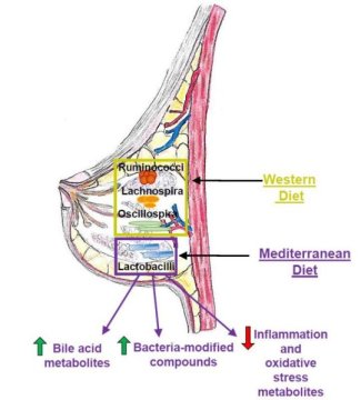
This image shows how diet plays a critical role in determining microbiota populations in tissues outside the gut, such as the mammary gland. Credit: Katherine Cook

"We showed for the first time that breast-specific microbiome populations are significantly affected by diet, and this was in a well-established nonhuman primate model of women's health, increasing the likelihood that these findings will be important for human health," says Carol Shively, Wake Forest School of Medicine. "The breast microbiome is now a target for intervention to protect women from breast cancer."
Diet has been extensively studied as a lifestyle factor that could influence breast cancer development. Breast cancer risk in women is increased by consumption of a high-fat Western diet full of sweets and processed foods but reduced by a healthy Mediterranean diet consisting of vegetables, fish, and olive oil. Intriguingly, a recent study in humans revealed that malignant breast tumors have a lower abundance of Lactobacillus bacteria compared to benign lesions, suggesting that microbial imbalances could contribute to breast cancer.
"However, it was unknown what possible factors could modulate the breast tissue microbiome," says Katherine Cook, Wake Forest School of Medicine. "Diet is a strong influencer on the gut microbiome, so we decided to test the hypothesis that diet can impact mammary gland microbiota populations."
To address this question, Shively and Cook used macaque monkeys because the animals mimic human breast biology and have been used to study breast cancer risk. One advantage over human studies is that the food intake of the monkeys can be carefully controlled for a prolonged period of time, increasing the chance of observing profound effects of diet.
The researchers assigned 40 adult female monkeys to receive either a Western or a Mediterranean diet for 31 months. The breast tissue of monkeys that consumed a Mediterranean diet had a 10-fold higher abundance of Lactobacillus, which is commonly used in probiotics and has been shown to decrease tumor growth in animals with breast cancer. The Mediterranean diet also increased levels of bile acid metabolites and bacterial-processed bioactive compounds that may decrease breast cancer risk.
Taken together, these results suggest that diet directly influences microbiome populations outside of the intestinal tract and could impact mammary gland health. But for now, it is not clear what impact these microbes or microbial-modified metabolites have on breast cancer risk.
Moving forward, the Cook lab plans to investigate the physiological impact of increased Lactobacillus in mammary gland tissue. They will also assess whether oral interventions such as fish oil or probiotic supplements can impact microbial populations in mammary glands and breast tumors. In addition, they are exploring the role of bacterial-modified bioactive compounds and bile acids on inflammation, breast cancer tumor growth, and therapeutic responsiveness.
"Our future studies are designed to validate the use of probiotics, fish oil, or antibiotics during neoadjuvant therapy to improve therapeutic outcomes," Cook says. "We hope to translate our studies into the clinic in the near future."
We encourage researchers all around the globe to submit abstract on their latest research at our upcoming conference Cell Tissue Science 2019 which is mainly focuses on the complications the consequences of Stem CellRegenerative MedicineStem Cell TherapyCancer Cell Biology , Technical Advancements in cancer treatment   and many more.We welcome you to the our upcoming conference “ 12th World Congress on Cell & Tissue Science” . For more info visit :Cell Tissue Science 2019