Showing posts with label Top Cancer Conferences. Show all posts

Saturday, 15 June 2019

'Virtual biopsy' device to detect skin tumors


Using sound vibrations and pulses of near-infrared light, a Rutgers University scientist has developed a new "virtual biopsy" device that can quickly determine a skin lesion's depth and potential malignancy without using a scalpel.

The ability to analyze a skin tumor non-invasively could make biopsies much less risky and distressing to patients, according to a report in Wiley Online Library. Currently, physicians who perform surgical biopsies often don't know the extent of a lesion and whether it will be necessary to refer the patient to a specialist for extensive tissue removal or plastic surgery until surgery has already begun.

The first-of-its-kind experimental procedure, called vibrational optical coherence tomography (VOCT), creates a 3-D map of the legion's width and depth under the skin with a tiny laser diode. It also uses soundwaves to test the lesion's density and stiffness since cancer cells are stiffer than healthy cells. An inch-long speaker applies audible soundwaves against the skin to measure the skin's vibrations and determine whether the lesion is malignant.
"This procedure can be completed in 15 minutes with no discomfort to the patient, who feels no sensation from the light or the nearly inaudible sound. It's a significant improvement over surgical biopsies, which are invasive, expensive and time consuming," said Frederick Silver, a professor of pathology and laboratory at Rutgers Robert Wood Johnson Medical School.
The study found that a prototype VOCT device, which awaits FDA approval for large-scale testing, is able to accurately distinguish between healthy skin and different types of skin lesions and carcinomas. The researchers tested the device over six months on four skin excisions and on eight volunteers without skin lesions. Further studies are needed to fine-tune the device's ability to identify a lesion's borders and areas of greatest density and stiffness, which would allow physicians to remove tumors with minimally invasive surgery.

An important announcement regarding our upcoming conference 12th World Congress on Cell & Tissue Science (Cell Tissue Science 2019) scheduled on September 13-14,2019 in Singapore. You can also present your latest research at the different topics such as Cancer Cell Biology, Stem Cell & its applications and many more along with other distinguished professors, doctors and researchers from all over the world.
If interested kindly proceed with submitting your abstract and latest biography along with a photography to our online abstract submission page given below: Link for submission: Click Here
Source: https://www.sciencedaily.com/releases/2019/06/190613103129.htm

Thursday, 24 January 2019

Energizing the immune system to eat cancer


Immune cells called macrophages are supposed to serve and protect, but cancer has found ways to put them to sleep. Now researchers at the Abramson Cancer Center of the University of Pennsylvania say they've identified how to fuel macrophages with the energy needed to attack and eat cancer cells. It is well established that macrophages can either support cancer cell growth and spread or hinder it. But most tumors also express a signal called CD47, which can lull macrophages into a deep sleep and prevent them from eating. Researchers have found that rewiring macrophage metabolism can overcome this signal and act like an alarm clock to rouse and prepare macrophages to go to work. Their findings were published in Nature Immunology today.

Macrophages are immune cells just like T and B cells, but differ in that they can eat cells that are not supposed to be in the body. In fact, they are the most prominent immune cell found in cancer, but unfortunately, most are often convinced to help cancer grow and spread. Cancer cells frequently stop macrophages from attacking them by expressing CD47, a "don't eat me" signal. Researchers now say that merely blocking inhibitory signals like CD47 is not always sufficient to convince macrophages to attack cancer. Instead, two signals are required. First, they need a signal to activate them -- such as a toll-like receptor agonist. After that, a second signal -- such as a CD47 inhibitor -- can lower the threshold needed to wage battle on the cancer.

"It turns out macrophages need to be primed before they can go to work, which explains why solid tumors may resist treatment with CD47 inhibitors alone," said the study's senior author Gregory L. Beatty, Assistant Professor of Hematology-Oncology at Penn's Perelman School of Medicine. 
The team used this approach by activating macrophages with CpG, a toll-like receptor agonist that sends the first signal, and found that it rapidly induced shrinkage of tumors and prolonged survival of mice even without the requirement of T cells. Unexpectedly, they also found that the activated macrophages were able to eat cancer cells even in the presence of high levels of CD47.

To understand the molecular basis of this phenomenon, the team traced the metabolic activity of macrophages and determined that activated macrophages began to utilize both glutamine and glucose as fuel to support the energy requirements needed for them to eat cancer cells. This rewiring of the macrophages metabolism was necessary for CpG to be effective, and the researchers say these findings point to the importance of macrophage metabolism in determining the outcome of an immune response.
"Cancer does not shrink without the help of macrophages and macrophages need the right fuel to eat cancer cells and shrink tumors," Liu said. "To do this, a shift in metabolism is needed to steer the energy in the right direction. It is the metabolism that ultimately allows macrophages to override signals telling them not to do their job."
Beatty points out that patients with diabetes, cardiovascular disease, and other conditions are routinely treated with drugs that could affect macrophage metabolism, but virtually nothing is known about how these drugs might impact immunotherapy responses in cancer, meaning the team's discovery has implications even for existing treatments.
Researchers from different part of the world are invited to submit abstract on their unpublished latest research at our upcoming conference Cell Tissue Science 2019 which is focused on the complications and consequences of Stem Cell, Regenerative Medicine, Stem Cell Therapy, Cancer Cell Biology,Technical Advancements in cancer treatment and many more. We as committee members of the conference welcome you to be a part of the conference “ 12th World Congress on Cell & Tissue Science” in Singapore on March 11-12, 2019. 
You can submit your abstract on Session or Track : 08. Advancement in Cancer Treatments

Wednesday, 23 January 2019

New nanoparticle targets tumor-infiltrating immune cells, flips switch


Immunotherapy's promise in the fight against cancer drew international attention after two scientists won a Nobel Prize this year for unleashing the ability of the immune system to eliminate tumor cells.

But their approach, which keeps cancer cells from shutting off the immune system's powerful T-cells before they can fight tumors, is just one way to use the body's natural defenses against deadly disease. A team of Vanderbilt University bioengineers today announced a major breakthrough in another: penetrating tumor-infiltrating immune cells and flipping on a switch that tells them to start fighting. The team designed a nanoscale particle to do that and found early success using it on human melanoma tissue.
"Tumors are pretty conniving and have evolved many ways to evade detection from our immune system," said John T. Wilson, Assistant Professor of Chemical and Biomolecular Engineering and Biomedical Engineering. "Our goal is to rearm the immune system with the tools it needs to destroy cancer cells.Checkpoint blockade has been a major breakthrough, but despite the huge impact it continues to have, we also know that there are a lot of patients who don't respond to these therapies. We've developed a nanoparticle to find tumors and deliver a specific type of molecule that's produced naturally by our bodies to fight off cancer."
That molecule is called cGAMP, and it's the primary way to switch on what's known as the stimulator of interferon genes (STING) pathway: a natural mechanism the body uses to mount an immune response that can fight viruses or bacteria or clear out malignant cells. Wilson said his team's nanoparticle delivers cGAMP in a way that jump-starts the immune response inside the tumor, resulting in the generation of T-cells that can destroy the tumor from the inside and also improve responses to checkpoint blockade.

While the Vanderbilt team's research focused on melanoma, their work also indicates that this could impact treatment of many cancers, Wilson said, including breast, kidney, head and neck, neuroblastoma, colorectal and lung cancer.

His findings appear today in a paper titled "Endosomolytic Polymersomes Increase the Activity of Cyclic Dinucleotide STING Agonists to Enhance Cancer Immunotherapy" in the journal Nature Nanotechnology.

Daniel Shae, a Ph.D. student on Wilson's team and first author of the manuscript, said the process began with developing the right nanoparticle, built using "smart" polymers that respond to changes in pH that he engineered to enhance the potency of cGAMP. After 20 or so iterations, the team found one that could deliver cGAMP and activate STING efficiently in mouse immune cells, then mouse tumors and eventually human tissue samples.
"That's really exciting because it demonstrates that, one day, this technology may have success in patients," Shae said.
Researchers from different part of the world are invited to submit abstract on their unpublished latest research at our upcoming conference Cell Tissue Science 2019 which is focused on the complications and consequences of Stem Cell, Regenerative Medicine, Stem Cell Therapy, Cancer Cell Biology,Technical Advancements in cancer treatment and many more. We as committee members of the conference welcome you to be a part of the conference “ 12th World Congress on Cell & Tissue Science” in Singapore on March 11-12, 2019. 
You can submit your abstract on Session or Track : 08. Advancement in Cancer Treatments

Monday, 21 January 2019

Why haven't cancer cells undergone genetic meltdowns?


Cancer first develops as a single cell going rogue, with mutations that trigger aggressive growth at all costs to the health of the organism. But if cancer cells were accumulating harmful mutations faster than they could be purged, wouldn't the population eventually die out?

How do cancer cells avoid complete genetic meltdown?
Famously isolated from cervical cancer victim Henrietta Lacks in 1951, they became the first immortalized cell line, helped in the development of the polio vaccine, and have become a biotechnology foundational resource for any in vitro drug development or cancer studies.

And they are still providing ample opportunities to further our understanding of cancer.
"In this study, HeLa cells are not used to reveal the process of tumorigenesis but mainly a model for addressing the underlying evolutionary forces, which need to be powerful enough to measure in laboratory settings. We examined variation in growth rate among individual HeLa cells by monitoring clones from a common ancestral HeLa cell population," said corresponding author Xuemei Lu.
They first established a HeLa cell line (E6) derived from an ancestral cell line. When the population size of E6 reached approximately 5 × 10⁴ cells (15~16 divisions), five single-cell clones were generated and established in culture. They team DNA sequenced these clones to catalog the mutations. They focused on copy number variation (CNV) rather than single DNA changes because single-nucleotide mutation rates are too slow to produce significant sequence variation during the short-duration culturing experiments.
"We then estimated the deleterious mutation rate and the average fitness decrease per mutation by performing computer simulations of cell growth," said author Hurng-Yi Wang.
Overall, they found that the main mutations affect the copy number of genes, with an average of 0.29 deleterious events for every cell division. Each of these events reduces fitness 18 percent.

Their results indicate that heterogeneity in cell growth can be generated in a very short period of time in cancer cells and is heritable and genetically determined.
"Our estimates indicate that the HeLa cells experience a 5 percent reduction (0.29 ×0.18 ? 5%) in fitness for every generation. Our observations suggest that human cells that have been cultured for a sufficiently long period still generate deleterious mutations in the form of CNVs at a high rate and with a high intensity. For such systems, a mutational meltdown might be plausible."
For example, when they isolated 39 cells from B8 (a fast-growing clone) and 40 cells from E3 (slow growing clone), and monitored their growth from a single cell for seven days, approximately 23 percent of B8 and 50 percent of E3 cells died out within seven days, due to either damage caused during cell isolation or genetic defects.

Most cell lines with growth rates < 0.6 died within 2 months. In total, only 60 percent of B8 and 27 percent of E3 cells survived for more than two months.

Next, they picked about 20 cells from each of the single cell originated clones from B8 and counted their chromosome numbers.

The chromosomes varied far from the normal human number of 46. They ranged from 38 to 113 chromosomes, with most (72 percent) cells harboring between 55 and 70 chromosomes, indicating that they are triploid. Therefore, despite single-cell origin, the progeny quickly generated aneuploidy within only 20-30 cell divisions, again illustrating frequent cytogenetic change in cancer cells.

Despite the level of mutations occurring, reduction in growth rates, and chromosome numbers no longer representing that of normal humans, cancer cells still find a way to survive.
So how do HeLa cells persist?
"High deleterious mutation rate would raise an impression that the HeLa cell lines may have gone extinct long ago," said Lu.
Their simulation results indicated that although most of the cells accumulated deleterious mutations and were worse than the ancestral cells, there were still 13.1 percent of cells which were mutation-free.

"These mutation-free cells can avoid the population from extinction."
It also explains why, even if chemotherapy treatment successfully killed 90 percent of a cancer cell population, it may still not be enough.

The new study not only advances the understanding of the evolution of HeLa cells, and of tumors in general, but of the cells of multicellular organisms in culture in general. In future work, the scientists want to exploit their cancer cell fitness and growth rate findings to understand how cancer cells can become even more vulnerable to recent breakthroughs with checkpoint inhibitor drugs.
Researchers from different part of the world are invited to submit abstract on their unpublished latest research at our upcoming conference Cell Tissue Science 2019 which is focused on the complications and consequences of Stem Cell, Regenerative Medicine, Stem Cell Therapy, Cancer Cell Biology,Technical Advancements in cancer treatment and many more. We as committee members of the conference welcome you to be a part of the conference “ 12th World Congress on Cell & Tissue Science” in Singapore on March 11-12, 2019. 
You can submit your abstract on Session or Track : 08. Advancement in Cancer Treatments
 

Thursday, 4 October 2018

Diet affects the breast microbiome in mammals

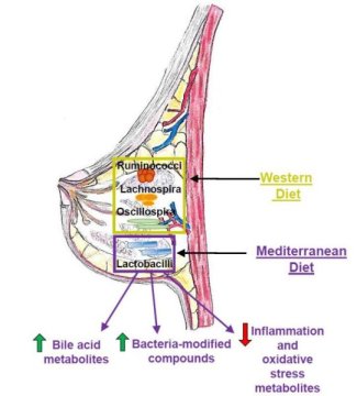
Diet influences the composition of microbial populations in the mammary glands of nonhuman primates. Specifically, a Mediterranean diet increased the abundance of probiotic bacteria previously shown to inhibit tumor growth in animals.
Cell Tissue Science 2019
This image shows how diet plays a critical role in determining microbiota populations in tissues outside the gut, such as the mammary gland. Credit: Katherine Cook

"We showed for the first time that breast-specific microbiome populations are significantly affected by diet, and this was in a well-established nonhuman primate model of women's health, increasing the likelihood that these findings will be important for human health," says Carol Shively, Wake Forest School of Medicine. "The breast microbiome is now a target for intervention to protect women from breast cancer."
Diet has been extensively studied as a lifestyle factor that could influence breast cancer development. Breast cancer risk in women is increased by consumption of a high-fat Western diet full of sweets and processed foods but reduced by a healthy Mediterranean diet consisting of vegetables, fish, and olive oil. Intriguingly, a recent study in humans revealed that malignant breast tumors have a lower abundance of Lactobacillus bacteria compared to benign lesions, suggesting that microbial imbalances could contribute to breast cancer.
"However, it was unknown what possible factors could modulate the breast tissue microbiome," says Katherine Cook, Wake Forest School of Medicine. "Diet is a strong influencer on the gut microbiome, so we decided to test the hypothesis that diet can impact mammary gland microbiota populations."
To address this question, Shively and Cook used macaque monkeys because the animals mimic human breast biology and have been used to study breast cancer risk. One advantage over human studies is that the food intake of the monkeys can be carefully controlled for a prolonged period of time, increasing the chance of observing profound effects of diet.
The researchers assigned 40 adult female monkeys to receive either a Western or a Mediterranean diet for 31 months. The breast tissue of monkeys that consumed a Mediterranean diet had a 10-fold higher abundance of Lactobacillus, which is commonly used in probiotics and has been shown to decrease tumor growth in animals with breast cancer. The Mediterranean diet also increased levels of bile acid metabolites and bacterial-processed bioactive compounds that may decrease breast cancer risk.
Taken together, these results suggest that diet directly influences microbiome populations outside of the intestinal tract and could impact mammary gland health. But for now, it is not clear what impact these microbes or microbial-modified metabolites have on breast cancer risk.
Moving forward, the Cook lab plans to investigate the physiological impact of increased Lactobacillus in mammary gland tissue. They will also assess whether oral interventions such as fish oil or probiotic supplements can impact microbial populations in mammary glands and breast tumors. In addition, they are exploring the role of bacterial-modified bioactive compounds and bile acids on inflammation, breast cancer tumor growth, and therapeutic responsiveness.
"Our future studies are designed to validate the use of probiotics, fish oil, or antibiotics during neoadjuvant therapy to improve therapeutic outcomes," Cook says. "We hope to translate our studies into the clinic in the near future."
We encourage researchers all around the globe to submit abstract on their latest research at our upcoming conference Cell Tissue Science 2019 which is mainly focuses on the complications the consequences of Stem Cell, Regenerative Medicine, Stem Cell Therapy, Cancer Cell Biology , Technical Advancements in cancer treatment   and many more.We welcome you to the our upcoming conference “ 12th World Congress on Cell & Tissue Science” . For more info visit :Cell Tissue Science 2019